
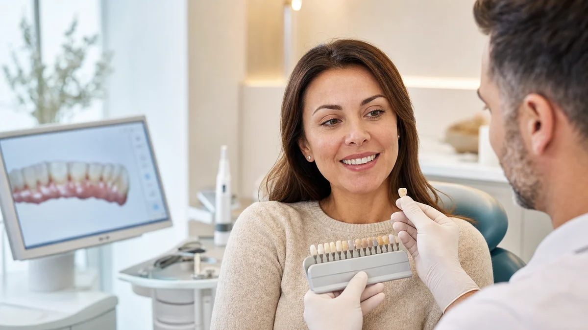
Quick answer: Dental crowns can restore shape, strength, colour and bite when a tooth is sufficiently damaged or heavily restored. A credible before-and-after assessment compares more than brightness: margins, symmetry, gum response, contact points and function matter. Results vary, and a clinical examination is essential before deciding whether a crown is appropriate.
Searches for dental crowns before and after often begin with photographs, but a photograph captures only one surface of a clinical decision. A crown is a custom restoration that covers a prepared tooth or sits on a dental implant. It may help protect a weakened tooth, rebuild a fractured one, restore a heavily filled tooth, or improve an unsuitable shape or colour. The American Dental Association’s patient information explains that crowns can strengthen teeth, cover badly shaped or discoloured teeth and restore implants. Those indications are different from simply making every visible tooth look identical.
A responsible review of dental crowns before and after should therefore ask two questions at once: does the smile look harmonious, and does the restoration support oral health and function? The answer depends on the condition of the tooth, the gums, the bite, the selected material, the laboratory design and the patient’s maintenance. This guide explains what can reasonably change, what cannot be promised and how to judge images and treatment plans without mistaking marketing for diagnosis.
Redent Klinik provides this information for education, not for remote diagnosis. If you are comparing options, begin with the Redent Klinik English homepage and arrange an individual assessment through the Redent Klinik contact page. Your dentist must examine the tooth and surrounding tissues before recommending irreversible preparation.
What dental crowns before and after images can genuinely show
Well-made dental crowns before and after records can document visible differences in tooth shape, edge position, colour, spacing and the balance of a smile. They can also show how a damaged or heavily restored tooth has been rebuilt. For front teeth, the comparison may reveal changes in width, length, surface texture and translucency. For back teeth, photographs are less informative because the most important changes may involve chewing anatomy, contact with neighbouring teeth and protection of the remaining tooth structure.
In dental crowns before and after reviews, images cannot prove that a crown fits accurately below or at the gum margin. They do not show whether decay was fully managed, whether the nerve is healthy, whether the bite is balanced or whether cleaning is easy. Nor do they tell you how the result feels months later. A clinically useful record may include photographs, radiographs when justified, digital scans, bite records and notes about symptoms. The visual comparison is one piece of that record, not a substitute for it.
- Visible changes: shape, apparent alignment, shade, surface character, chips and gaps.
- Functional changes: contact points, bite balance, speech adaptation and chewing comfort.
- Biological considerations: gum health, remaining tooth structure, decay risk and pulp health.
- Maintenance factors: floss access, plaque control, grinding habits and review attendance.
The best dental crowns before and after discussion connects these four areas. A crown that looks bright in a photograph but causes persistent pain, traps food or inflames the gum is not a successful outcome. Conversely, a carefully fitted molar crown may create little visible drama while delivering meaningful protection and function.
12 realistic changes to compare before and after a crown
Every tooth is different, so this is not a checklist of guaranteed outcomes. It is a framework for discussing dental crowns before and after with your dentist. Some changes are visible immediately, while others need follow-up and professional assessment.
- Overall tooth shape: a fractured, worn or irregular outline may be rebuilt to a planned contour.
- Edge position: a front crown can alter the length and alignment of the visible incisal edge.
- Shade integration: the restoration can be matched to adjacent teeth, but exact perception varies with lighting.
- Surface texture: subtle ridges and gloss can help a crown reflect light more naturally.
- Translucency: ceramic systems can imitate aspects of enamel, although materials behave differently.
- Visible old restorations: dark, broken or oversized fillings may be covered after appropriate assessment and preparation.
- Contact with neighbours: a properly designed contact may reduce food packing without making floss impossible.
- Bite contact: the new surface must meet opposing teeth in a controlled way during closing and movement.
- Gum contour: healthy tissue may frame the crown naturally, but gum position is not completely controlled by the restoration.
- Speech and lip support: front-tooth contours can influence sounds and how the lip rests, especially when several teeth are treated.
- Cleaning access: the final contour should allow daily plaque removal around the margin.
- Confidence: some people feel more comfortable smiling, but emotional outcomes are personal and cannot be guaranteed.
When reviewing dental crowns before and after, ask which of these changes was a treatment goal and how it was measured. A single frontal photograph may support discussion of shade and shape, but it cannot establish the quality of the bite, internal tooth health or margin adaptation.
What happens before the “before” photograph
In dental crowns before and after planning, the visible starting point is only the beginning. Before recommending a crown, a dentist usually asks about symptoms, dental history, medical conditions, medicines and expectations. The tooth and gums are examined. Depending on the clinical question, radiographs or vitality tests may be considered. Existing decay, cracks, large fillings, root canal history and the amount of sound tooth remaining all affect the plan.
For meaningful dental crowns before and after planning, the dentist also assesses the bite. Clenching or grinding can place high loads on ceramics and tooth structure. A deep bite, limited space or an unfavourable contact pattern may change the preparation, material or protective plan. Gum inflammation should be addressed because swollen or bleeding tissue makes accurate impressions and predictable margins more difficult.
Photographs used for dental crowns before and after should be standardised when they are used for comparison. The camera angle, facial position, lens distance, lighting, retraction, tooth hydration and colour calibration can all influence what viewers perceive. Teeth temporarily become lighter when dehydrated during a long appointment, then regain their usual appearance after rehydration. This is why a dramatic online comparison may not represent the same conditions on both sides.
- Ask what problem the crown is intended to solve.
- Ask whether less invasive options are clinically reasonable.
- Discuss gum health, decay risk and grinding or clenching.
- Review the proposed shade and shape in more than one light source when possible.
- Clarify whether treatment involves a natural tooth, a root-treated tooth or an implant.
How a dental crown is prepared and fitted
A typical dental crowns before and after pathway starts with preparation. The NHS describes a crown as a cap that completely covers a real tooth and notes that the tooth must be shaped so the crown can be fixed. The exact sequence varies with the tooth, material and clinical situation. Local anaesthesia may be used. Damaged or decayed tissue is managed, and the tooth is prepared to create enough space for the chosen restoration while preserving appropriate structure. A build-up may be needed if substantial tissue is missing.
For complete dental crowns before and after records, the dentist may document the prepared tooth and nearby teeth using a digital scan or conventional impression. Bite information and shade details are sent to a dental laboratory or used in an in-clinic digital workflow. In many laboratory cases, a temporary crown protects the tooth while the definitive restoration is made. A temporary is not expected to reproduce every final detail, and it may require more cautious cleaning and eating.
At the fit appointment, the dentist checks the crown’s seating, margins, contact points, shade and bite before final cementation or bonding. Adjustments may be necessary. The “after” moment in dental crowns before and after is therefore not merely the instant a bright shell appears; it follows a sequence of diagnostic, technical and biological checks. Some cases need additional review after the gums settle or after the patient has used the new bite.
A crown preparation is generally irreversible because tooth structure is altered. That fact should be part of every dental crowns before and after consent discussion. A dentist should explain reasonable alternatives, material-related trade-offs, expected maintenance and case-specific risks. The safest plan is the one supported by examination rather than a plan copied from another person’s photographs.
Choosing a material: appearance is only one criterion
Material choice changes how dental crowns before and after may look and function. Dental crowns may be made from ceramic, zirconia, porcelain fused to metal, metal alloys or resin-based materials used in selected situations. No material is universally best. Location, available space, bite force, aesthetic priority, the condition of the supporting tooth, allergies and the design of the opposing teeth all matter. The laboratory’s skill and the quality of tooth preparation and cementation also influence the result.
In anterior dental crowns before and after, all-ceramic restorations are often selected for their ability to reproduce colour, fluorescence and translucency. However, a very dark underlying tooth or metal post may influence the optical plan. More opaque ceramics can mask colour but may need careful layering or staining to avoid a flat appearance. A natural smile is not a row of identical white rectangles; small variations in value, texture and edge form create realism.
In posterior dental crowns before and after, strength, thickness, space and wear against opposing enamel become particularly important. A highly polished, well-designed surface behaves differently from a rough or over-contoured one. Material names alone do not predict success. Ask why a material is suitable for your tooth and what maintenance it requires.
| Decision factor | What to discuss | Why it affects the result |
|---|---|---|
| Front or back tooth | Visibility, load and available restorative space | Optical priorities and chewing forces differ by position |
| Remaining tooth structure | Cracks, old fillings, decay and retention | The foundation influences whether a crown is appropriate and how it is prepared |
| Shade and substrate | Adjacent tooth colour and the colour beneath the crown | Opacity and translucency must be planned together |
| Bite and grinding | Contact pattern, wear signs and possible night guard | High or repeated forces increase complication risk |
| Gum and decay risk | Inflammation, dry mouth, hygiene and diet | Margins remain vulnerable to plaque-related disease |
| Aesthetic expectation | Natural integration versus a deliberately brighter change | A shared target reduces misunderstandings about the “after” appearance |
This decision table helps make dental crowns before and after discussions patient-specific. It does not select a material on its own. Your dentist and dental technician must combine examination findings with your informed preferences.
How to read shade, shape and symmetry in photographs
Colour interpretation is central to dental crowns before and after. Value describes relative lightness, chroma describes intensity and hue describes the colour family. Translucency, surface gloss and the surrounding lips and skin also affect perception. A tooth photographed under a cool ring light can look different from the same tooth in warm daylight. Image editing, exposure and white balance can intensify that effect.
When comparing dental crowns before and after, look for integration rather than maximum whiteness. Does the crown reflect light similarly to adjacent teeth? Is the surface too uniform? Are the edges compatible with the patient’s age and neighbouring teeth? Does the midline look balanced within the face rather than merely centred in a cropped mouth? Symmetry can be desirable, but perfect mirror-image teeth may appear artificial.
Shape in dental crowns before and after must also respect biology. An over-contoured crown may look full in a frontal view but make plaque control difficult near the gum. An under-contoured contact area can trap food. A crown that is too long or bulky can affect speech, lip movement or bite. These relationships require clinical examination and side views; they cannot be judged from a single carefully selected frame.
Ethical dental crowns before and after galleries should use comparable angles and lighting and should not imply that another person will receive the same result. Patient consent and privacy matter. Images are educational examples, not warranties.
Gums, margins and the hidden part of the result
The hidden margin is essential when assessing dental crowns before and after. This junction between crown and tooth has a design and location that depend on the tooth, restoration and clinical need. The margin should be accurately finished and accessible to professional assessment and daily plaque control. Photographs may show the visible gum line, but they cannot reveal every part of the interface.
Healthy gums are part of a stable dental crowns before and after result. They generally appear firm and comfortable and should not bleed repeatedly with gentle cleaning. Temporary irritation may occur after treatment, but persistent swelling, bleeding, bad taste, discharge or increasing discomfort needs dental review. Gum recession can expose the crown margin or root surface over time. Tissue position may change with inflammation control, ageing, brushing trauma, periodontal disease or tooth movement.
This is a key limitation of dental crowns before and after: the immediate photograph may be taken before tissue maturation. A review several weeks or months later can provide different information. The biological “after” continues beyond cementation, and maintenance is part of the restoration’s future.
Long-term dental crowns before and after care recognises that a crown does not make the underlying tooth immune to decay. New decay can develop at an exposed or plaque-retentive margin. Daily fluoride toothpaste, cleaning between teeth, a balanced diet and risk-based dental reviews remain important. The World Health Organization emphasises twice-daily brushing with fluoride toothpaste and reducing free-sugar exposure as core preventive measures.
Recovery, sensitivity and warning signs after placement
Recovery is another part of dental crowns before and after. Mild gum tenderness or temperature sensitivity can occur after preparation or cementation, depending on the tooth and procedure. It should generally improve rather than intensify. The bite may feel unfamiliar at first, but a crown that feels clearly high, prevents normal closure or causes pain on biting should be checked. Do not wait for severe symptoms in the hope that the restoration will “wear in.”
During the temporary phase, follow the clinic’s cleaning and eating instructions. Very sticky or hard foods can dislodge or fracture some temporary restorations. If a temporary crown comes off, contact the dental team; prolonged exposure may allow sensitivity or tooth movement that complicates the final fit.
After final placement, seek timely dental advice for persistent or increasing pain, swelling, fever, a loose crown, a new crack, sharp edges, a bad taste or difficulty biting. Urgent assessment is appropriate when facial swelling affects breathing or swallowing, or when severe symptoms escalate rapidly. Online dental crowns before and after examples cannot triage these problems.
- Use a soft-bristled toothbrush and fluoride toothpaste as advised.
- Clean between the crown and neighbouring teeth every day with a suitable method.
- Avoid using crowned teeth to open packaging or bite non-food objects.
- Discuss a protective appliance if you clench or grind.
- Attend reviews based on your dentist’s assessment of decay and gum risk.
Crowns, veneers, fillings and onlays: why the alternatives matter
A crown covers most or all of the prepared clinical tooth. A veneer usually covers the front surface and is mainly considered for selected aesthetic changes when the underlying tooth is suitable. A filling replaces a smaller area of lost structure. An inlay or onlay can restore a portion of the chewing surface and cusps in suitable cases. Bonding may address limited shape or colour concerns with less extensive preparation, although it has different maintenance and durability characteristics.
The most impressive dental crowns before and after transformation is not necessarily the most conservative solution. If a tooth is largely healthy and the concern is minor, extensive preparation may not be justified. If a tooth is severely cracked or heavily restored, a small filling may not provide adequate coverage. Only an examination can locate a patient on that spectrum.
Root canal treatment is not automatically required for every crown. It may be indicated when the pulp is irreversibly inflamed, infected or already treated for another reason. Likewise, not every root-treated tooth receives the same type of restoration. Tooth location, remaining structure and loading influence the plan. These distinctions are important because oversimplified galleries can make a complex pathway appear like one cosmetic appointment.
Ask the dentist to explain the least invasive predictable options, what tooth structure each option removes, the likely maintenance and what could happen if treatment is postponed. Informed consent includes the option of no immediate treatment when clinically reasonable, together with the risks of monitoring.
How long the “after” result may last
No crown has a guaranteed lifespan. Survival depends on the supporting tooth, margin quality, decay risk, bite forces, material, cementation, gum health, trauma and daily care. A crown may require repair, recementation, root canal treatment or replacement if complications arise. The tooth itself can also fracture or develop disease independently of the crown material.
Long-term dental crowns before and after records are more informative than same-day photographs because they can show tissue response, wear, shade stability and maintenance. Even then, another patient’s time course cannot predict yours. Statements that a restoration will last a fixed number of years should be treated as general estimates, not promises.
Regular monitoring aims to identify problems while they are more manageable. A dentist may assess margins, contacts, bite, gum measurements, symptoms and radiographic findings when indicated. At home, watch for floss catching or shredding, recurrent food trapping, new sensitivity, movement, chips or changes in the surrounding gum. Early contact with the dental team is preferable to waiting for visible failure.
Aesthetic preferences can also change. Replacing a healthy crown only to obtain a different shade exposes the tooth to another procedure and must be weighed carefully. Whitening natural teeth after crown placement will not whiten the crown in the same way, so sequence and shade planning matter before treatment.
Questions to ask at a crown consultation
Bring your priorities to the consultation, but allow the examination to shape the plan. If you have saved dental crowns before and after images, explain what you like about them: perhaps the natural edge shape, reduced dark space or softer colour. The images can support communication without becoming a demand for an identical copy.
- What diagnosis or structural problem makes a crown appropriate for this tooth?
- What alternatives are reasonable, including monitoring, a filling, an onlay or another restoration?
- How much tooth preparation is expected, and is the process reversible?
- Which material is proposed, and why does it suit my bite, tooth and aesthetic goal?
- Will I need a temporary crown, and how should I care for it?
- What are the case-specific risks to the nerve, gum, bite and restoration?
- How will shade, contact points, margins and bite be checked before final cementation?
- What symptoms should prompt a routine, prompt or urgent review?
- How will grinding, dry mouth, gum disease or high decay risk affect maintenance?
- What records will be used to assess the result over time?
A clear answer about dental crowns before and after does not have to promise perfection. It should connect the proposed restoration to your clinical findings and explain uncertainty. That is a stronger basis for consent than a gallery alone.
Frequently asked questions about dental crowns before and after
Do dental crowns before and after photos predict my result?
No. Dental crowns before and after photos can illustrate possibilities and help you describe preferences, but they cannot predict your anatomy, tissue response, shade match or healing. Camera settings and editing also affect appearance. Your dentist must assess your tooth structure, gums, bite and expectations before providing an individual plan.
Will a crown look exactly like a natural tooth?
A carefully designed crown can imitate many features of enamel, including colour gradients, surface texture and translucency. Exact invisibility cannot be guaranteed. The underlying tooth colour, material thickness, lighting, neighbouring restorations and gum position all affect perception. In dental crowns before and after records, a natural integration is usually a more useful goal than maximum whiteness.
Can my teeth be whitened after a crown is fitted?
Whitening products primarily change natural tooth colour and do not predictably lighten crown materials in the same way. This can create a mismatch. If whitening is appropriate, it is often discussed before the definitive crown shade is selected, with time allowed for colour stabilisation. A dentist should assess sensitivity, decay and gum health first.
Is sensitivity normal after a dental crown?
Some people experience short-term temperature sensitivity or gum tenderness. It should trend toward improvement. Persistent, severe or worsening pain, swelling, pain that disturbs sleep, or pain on biting needs assessment. A high bite, pulp inflammation, cement irritation, a crack or another condition may require professional investigation rather than self-diagnosis.
Why does my new crown feel too high?
Even a small premature contact may make a restoration feel prominent. The sensation should be evaluated if normal closure or chewing feels altered, especially when pain develops. The dentist can assess contacts with clinical methods and adjust when appropriate. Do not deliberately grind the crown down yourself or rely on prolonged discomfort to correct it.
Can decay develop under or around a crown?
Yes. The crown material itself does not decay, but the tooth remains vulnerable near the margin, particularly when plaque accumulates or dry mouth and frequent sugar exposure raise risk. Daily fluoride toothpaste, interdental cleaning and professional reviews remain necessary. New symptoms or a visible margin change should be examined.
How should I compare dental crowns before and after fairly?
Compare similar angles, lighting, lip position and tooth hydration. Look beyond colour to shape, gum response and harmony with neighbouring teeth. Ask when the final image was taken and whether the patient had other procedures. The most reliable dental crowns before and after evaluation also includes bite, margin and maintenance findings that photographs cannot show.
When should a crown be replaced?
Replacement may be considered when there is recurrent decay, an unrepairable fracture, a persistent fit problem, significant wear, loss of retention or another clinical concern. Age alone is not a diagnosis. Removing a crown is another intervention, so the dentist should weigh benefits, risks and alternatives after examination and, when indicated, imaging.
Are dental crowns only cosmetic?
No. Crowns are frequently restorative: they may protect or rebuild teeth weakened by fracture, wear, large restorations or other damage, and they can restore dental implants. Aesthetic improvement may accompany treatment, especially for visible teeth, but the indication should be connected to the health, structure and function of the individual tooth.
A safer way to make your decision
The central lesson from dental crowns before and after is that appearance and health must be assessed together. A crown can create meaningful improvement when the diagnosis, preparation, material, laboratory work, fitting and maintenance align. It is not a universal answer for every discoloured, crowded or imperfect tooth, and it cannot make oral tissues maintenance-free.
Use photographs to clarify your preferences, then ask for an examination-based explanation of the problem, alternatives and trade-offs. Make sure you understand that tooth preparation is irreversible, that complications are possible and that outcomes vary. A thoughtful plan should protect sound tissue where possible, produce cleansable contours and establish a bite that can be monitored.
If you are considering treatment at Redent Klinik, the next step is an individual consultation rather than a promise based on someone else’s image. Bring relevant records and a list of medicines, describe any pain or sensitivity accurately, and discuss your desired level of change. That process turns dental crowns before and after from a marketing phrase into a structured, patient-centred conversation.
Evidence and patient-information sources
- American Dental Association MouthHealthy: Crowns — patient information on why crowns may be recommended.
- American Dental Association — professional organisation and evidence-based dentistry resources.
- NHS: Dental treatments and crowns — overview of crown materials, preparation and temporary restorations.
- World Health Organization: Oral health fact sheet — prevention, fluoride toothpaste and oral-disease context.
Sources were checked on 22 July 2026. This article provides general education and does not replace diagnosis, examination or personalised consent.