
In 2025, understanding what a dental x ray is and why it is crucial for oral health has never been more important. A dental x ray is a diagnostic imaging tool used by dentists to visualize the teeth, gums, jawbone, and surrounding structures that are not visible during a standard dental examination. These high-resolution images help detect cavities, bone loss, infections, impacted teeth, and even early signs of oral cancer. Modern technology ensures that today’s dental x ray procedures are quick, painless, and expose patients to minimal radiation levels, making them safe for adults and children alike.
The dental x ray process uses low-dose radiation to capture detailed images of your oral cavity. Depending on the situation, your dentist might recommend bitewing, periapical, panoramic, or cone-beam computed tomography (CBCT) scans. Each type of dental x ray provides unique insights — from spotting a tiny cavity between molars to evaluating bone density before a dental implant procedure. With accurate imaging, treatment plans become more precise, reducing complications and ensuring long-term dental health.
One of the biggest advantages of a dental x ray is its ability to detect issues early. For example, cavities between teeth can develop without any pain or visible signs. Without an x-ray, these can progress to more severe conditions, such as root infections or gum disease. Similarly, impacted wisdom teeth, cysts, and jawbone irregularities often remain unnoticed without imaging. By incorporating a dental x ray into routine checkups, dentists can catch problems before they escalate, potentially saving patients from costly and invasive treatments.
Some patients worry about radiation exposure from a dental x ray. However, the amount of radiation is extremely low — often less than a day’s worth of natural background exposure. In fact, advancements in digital radiography have reduced radiation levels by up to 90% compared to traditional film x-rays. Dentists also use protective lead aprons and thyroid collars to further minimize exposure, especially for pregnant patients and children.
In addition to detecting dental issues, a dental x ray plays a role in planning various treatments. Orthodontists use them to determine tooth alignment and jaw position before recommending braces or Invisalign. Oral surgeons rely on them before extractions or implant placements. Even general dentists use them to monitor tooth root development in children, ensuring proper growth and spacing. By providing a complete view of oral structures, a dental x ray helps ensure safe and successful outcomes.
For patients in different parts of the world, the availability and cost of a dental x ray can vary. In the United States, a single bitewing x-ray might cost between $25 and $50, while a full-mouth series can range from $100 to $250. In the UK, NHS patients may pay as part of a treatment band fee, while private clinics may charge between £20 and £50 per image. In Turkey, dental x ray services are often more affordable, with prices starting as low as $15 for basic scans — making it a popular destination for dental tourism.
If you’re planning to get a dental x ray soon, it’s a good idea to choose a clinic that uses modern digital imaging systems. These not only produce clearer images but also allow your dentist to share them with specialists instantly. For patients looking for expert dental care in Turkey, you can Redent Klinik Contact Page to book your appointment. For general oral health guidelines, you can visit the American Dental Association website, which offers comprehensive resources.
Ultimately, a dental x ray is a vital tool in preventive dentistry. It empowers both patients and dentists with the knowledge needed to maintain healthy teeth and gums. In 2025, with rapid advances in dental technology, getting a dental x ray is safer, faster, and more accessible than ever before — ensuring brighter smiles and healthier lives for everyone. 😊
Understanding What a Dental X Ray Is and How It Works
A dental x ray is an essential diagnostic tool in modern dentistry, allowing professionals to see beneath the surface of your teeth and gums. While a visual examination can reveal visible issues such as cavities or plaque buildup, a dental x ray goes much deeper, capturing detailed images of tooth roots, jawbone structure, and areas between teeth that are otherwise hidden from view. This capability makes the dental x ray indispensable for early detection and effective treatment planning.
The technology behind a dental x ray involves the use of controlled radiation to penetrate oral tissues and produce an image on either digital sensors or traditional film. In digital radiography — the most common method in 2025 — the sensor instantly transmits the image to a computer screen, enabling immediate analysis. Traditional film-based x-rays, while still used in some clinics, require chemical processing and are generally less efficient. Digital dental x ray systems not only produce higher-quality images but also reduce radiation exposure significantly.
When a dental x ray is taken, the x-ray machine emits a focused beam of radiation that passes through the mouth. Dense materials like teeth and bones absorb more radiation and appear white on the image, while less dense tissues such as gums appear darker. This contrast helps dentists identify abnormalities such as tooth decay, infections, bone loss, impacted teeth, or developmental issues in growing children. It’s worth noting that the process is completely painless and usually takes less than a minute per image.
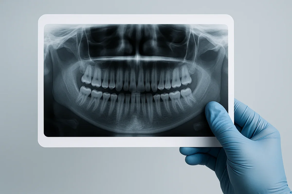
There are several types of dental x ray images, each serving a unique purpose. Bitewing x-rays capture the upper and lower teeth in one image, making them ideal for detecting cavities between teeth. Periapical x-rays show the entire tooth from crown to root tip, useful for diagnosing root infections. Panoramic x-rays provide a full view of the mouth, jaw, and sinuses, often used before orthodontic treatment or oral surgery. Cone-Beam Computed Tomography (CBCT) scans create 3D images, offering unparalleled detail for complex cases such as implant planning or detecting hidden fractures.
One of the key benefits of a dental x ray is its role in preventive care. Even if you have no visible symptoms, these images can reveal problems in their earliest stages, allowing for quicker and less invasive treatments. For instance, a minor cavity spotted on a dental x ray can be treated with a small filling, while the same cavity left undetected might later require root canal therapy. This early detection not only saves money but also helps preserve natural tooth structure.
Patients sometimes express concern about radiation exposure from a dental x ray. However, the levels are extremely low and comparable to the natural background radiation received in a single day. Digital radiography further reduces exposure, and protective equipment such as lead aprons and thyroid collars add an extra layer of safety. Pregnant women should inform their dentist before undergoing a dental x ray, although modern techniques are generally considered safe during pregnancy.
For those seeking advanced dental care in Turkey, choosing a clinic with state-of-the-art imaging equipment ensures accurate diagnoses and effective treatment plans. Facilities like Redent Klinik Contact Page offer modern dental x ray services with minimal radiation and high-resolution results. Additionally, the American Dental Association provides valuable information on the importance of x-rays in dental health.
Understanding how a dental x ray works helps patients appreciate its importance. It’s not just about detecting problems — it’s about preventing them, ensuring your smile stays healthy and beautiful for years to come. With today’s digital advancements, getting a dental x ray is faster, safer, and more accurate than ever, making it a cornerstone of 21st-century dental care. 🦷
Different Types of Dental X Rays and Their Uses
In 2025, there are multiple types of dental x ray techniques available, each designed to capture specific details of your oral health. Understanding these variations helps patients know what to expect during their dental visits and why a dentist might choose one type over another. Every dental x ray serves a unique diagnostic purpose, whether it’s identifying early tooth decay, assessing jaw alignment, or planning complex procedures like dental implants.
1. Bitewing X Rays – A bitewing dental x ray captures images of the upper and lower teeth in one area of the mouth. This type is particularly effective for spotting cavities between teeth and monitoring bone levels to detect gum disease. Dentists usually recommend bitewings once or twice a year as part of routine check-ups.
2. Periapical X Rays – A periapical dental x ray focuses on one or two teeth at a time, showing the crown, root, and surrounding bone. It is essential for diagnosing root infections, detecting cysts, and evaluating bone health before treatments like root canals.
3. Panoramic X Rays – A panoramic dental x ray offers a broad view of the entire mouth, including all teeth, both jaws, sinuses, and surrounding structures. This is often used before orthodontic work, wisdom tooth extractions, or implant surgery. It’s non-invasive and quick, usually completed in under a minute.
4. Occlusal X Rays – This type of dental x ray is taken with the mouth closed, capturing the bite of the upper or lower jaw. It helps detect jaw fractures, cysts, impacted teeth, and other structural issues.
5. Cone-Beam Computed Tomography (CBCT) – The CBCT dental x ray produces 3D images, offering exceptional detail for complex dental cases. Dentists use it for implant planning, evaluating jawbone density, or detecting hidden anatomical variations. While it emits more radiation than standard x-rays, it provides crucial information for surgical precision.
Choosing the right type of dental x ray depends on the patient’s needs. For instance, a routine cleaning appointment may only require bitewings, while a dental implant consultation might need a CBCT scan. Some treatments may combine multiple types of dental x ray imaging to ensure the most accurate diagnosis and treatment planning.
Advances in imaging technology mean that today’s dental x ray machines produce clearer images with lower radiation doses than ever before. Digital sensors have largely replaced film, enabling dentists to store, share, and enhance images instantly. This improves communication between specialists, speeds up treatment planning, and ensures patient comfort.
For patients considering dental tourism, countries like Turkey offer affordable and high-quality imaging services. Clinics such as Redent Klinik Contact Page provide advanced dental x ray technology comparable to top clinics worldwide. Meanwhile, resources like the American Dental Association explain the role of each x-ray type in maintaining oral health.
By understanding the different types of dental x ray procedures, you can make informed decisions about your care. Whether it’s a simple cavity check or a complex surgical plan, the right imaging method ensures accurate diagnosis, safe treatment, and a healthier smile. 🦷
Why Dental X Rays Are Important for Oral Health
A dental x ray is far more than just a diagnostic image—it’s a crucial component of preventive dentistry. By revealing hidden issues that cannot be detected during a routine visual examination, a dental x ray allows dentists to take early action, preventing minor problems from becoming major dental health concerns. Without this imaging tool, conditions such as cavities between teeth, bone loss, or infections in the tooth root could go unnoticed until they cause significant pain or damage.
One of the most significant benefits of a dental x ray is early detection. For example, gum disease often develops silently, with no immediate symptoms. A dental x ray can show bone loss in its earliest stages, enabling timely intervention that can save teeth and prevent further deterioration. Similarly, small cavities hidden between teeth are nearly impossible to spot without x-rays, yet catching them early can mean the difference between a simple filling and a more invasive procedure like a root canal.
A dental x ray also plays a key role in treatment planning. Whether you need orthodontic braces, dental implants, or wisdom tooth extraction, accurate imaging ensures that your dentist or specialist understands the exact positioning of your teeth, jawbone, and surrounding structures. This precision reduces surgical risks and improves the overall success rate of complex procedures.
Another important function of a dental x ray is monitoring changes over time. Dentists often compare current images to previous ones, tracking the progression of conditions such as bone density loss or the healing process after oral surgery. This long-term view helps in adjusting treatment plans and ensuring optimal results.
For children, a dental x ray is invaluable in monitoring tooth and jaw development. Pediatric dentists use it to check if permanent teeth are erupting correctly, if baby teeth are falling out on schedule, and if there are any developmental issues that may require early intervention. This proactive approach helps ensure that young patients grow up with healthy and well-aligned smiles.
Patients sometimes hesitate to get a dental x ray due to concerns about radiation exposure. However, as mentioned in other sections, modern digital x-rays emit minimal radiation—often less than what you receive from natural background sources in a single day. Furthermore, dentists follow strict safety protocols, using protective gear and only taking x-rays when necessary for diagnosis or monitoring.
Without the insight provided by a dental x ray, dentists would be working with limited information. This could lead to missed diagnoses, delayed treatments, and increased costs for the patient in the long run. By making a dental x ray part of your regular dental check-up, you ensure that potential problems are identified and addressed promptly, keeping your teeth and gums healthy for years to come.
For patients seeking professional care in Turkey, advanced imaging services are widely available. Clinics like Redent Klinik Contact Page combine high-quality dental x ray technology with expert dental care, ensuring accurate diagnoses and effective treatment plans. You can also find comprehensive oral health information on the American Dental Association website.
Ultimately, the importance of a dental x ray lies in its ability to safeguard your oral health. By catching problems early, guiding treatment decisions, and tracking changes over time, it plays a central role in maintaining a healthy, confident smile. 😊
Dental X Ray Procedure: Step-by-Step Process
For many patients, the thought of getting a dental x ray can cause a little uncertainty. However, the process is quick, safe, and entirely painless. Understanding each step of the dental x ray procedure can help you feel more comfortable and prepared for your appointment.
Step 1 – Initial Consultation
Before taking a dental x ray, your dentist will review your medical and dental history. They’ll ask about any symptoms you’re experiencing, such as pain, sensitivity, or swelling. This helps determine which type of dental x ray is most appropriate for your needs. Pregnant patients should inform their dentist so that extra safety precautions can be taken.
Step 2 – Preparation
You may be given a lead apron to wear during your dental x ray. This protective garment shields your body from minimal radiation exposure. In some cases, a thyroid collar may also be used to protect your neck area. These safety measures are standard in modern dental clinics worldwide.
Step 3 – Positioning
Depending on the type of dental x ray being taken, you will either sit in a dental chair or stand near the x-ray machine. The technician will position the sensor or film inside your mouth, ensuring it is comfortably placed to capture the required area. For panoramic x-rays, the machine will rotate around your head while you remain still.
Step 4 – Image Capture
Once you’re in position, the technician will step out of the immediate area to activate the x-ray machine. The process only takes a few seconds. You may hear a faint click or beep as the dental x ray is captured. For digital radiography, the image appears instantly on the computer screen for review.
Step 5 – Review of Results
Your dentist will analyze the dental x ray images, looking for signs of cavities, infections, bone loss, impacted teeth, or other abnormalities. They may zoom in on certain areas for a closer inspection. If necessary, the dentist will explain the findings and recommend a treatment plan.
Step 6 – Follow-Up
If your dental x ray reveals issues that require treatment, you may be scheduled for further procedures. In some cases, your dentist might request additional imaging for a more detailed view, especially if complex dental work is planned.
One advantage of modern dental x ray systems is their speed and efficiency. The entire process, from preparation to image review, usually takes less than 10 minutes. This makes it easy to incorporate into routine check-ups without causing significant delays in your dental visit.
Clinics that invest in the latest imaging technology, like Redent Klinik Contact Page, can offer a more comfortable experience with clearer images and reduced radiation exposure. Additionally, resources such as the American Dental Association provide guidelines on when and how often a dental x ray should be taken for different patient needs.
By understanding the step-by-step process, patients can approach a dental x ray appointment with confidence. Whether it’s a simple bitewing check or a full panoramic scan, this quick and straightforward procedure is a key part of maintaining excellent oral health. 🦷
Safety of Dental X Rays: Myths vs. Facts
The safety of a dental x ray is a common topic among patients, with many holding misconceptions about radiation exposure. While it’s natural to be cautious, modern dental x ray technology has made the process exceptionally safe, with minimal risks and significant benefits for oral health. In this section, we’ll separate myths from facts so you can make informed decisions about your dental care.
Myth 1 – Dental X Rays Expose You to Dangerous Radiation
Fact: The radiation from a single dental x ray is extremely low. In fact, a routine bitewing x-ray exposes you to less radiation than you’d naturally receive in a day from the environment. Digital x-ray systems, now widely used in 2025, reduce exposure by up to 90% compared to traditional film-based methods.
Myth 2 – Dental X Rays Are Not Necessary Unless You Have Pain
Fact: A dental x ray can reveal issues long before pain or visible symptoms appear. Cavities between teeth, bone loss, and impacted teeth are often invisible to the naked eye but are easily detected through x-rays. Early detection means less invasive treatment and better long-term results.
Myth 3 – Pregnant Women Should Never Get a Dental X Ray
Fact: While it’s important to limit radiation during pregnancy, modern dental x ray procedures are generally safe with proper shielding. Dentists use lead aprons and thyroid collars to protect both mother and baby. However, non-urgent x-rays may be postponed until after delivery as a precaution.
Myth 4 – Frequent Dental X Rays Are Harmful
Fact: Dentists follow strict guidelines to ensure x-rays are taken only when necessary. The frequency depends on factors like age, dental history, and current oral health. For most adults, a dental x ray is recommended once every 1–2 years unless specific issues require closer monitoring.
Myth 5 – All Dental X Rays Are the Same
Fact: There are various types of dental x ray images, each designed for specific purposes. Some, like bitewings, are for detecting cavities, while panoramic or CBCT scans provide detailed views for surgical planning. Your dentist selects the type based on your individual needs.
Safety protocols are a standard part of every dental x ray procedure. These include using the lowest radiation dose possible, proper patient positioning, and protective equipment. Technological advances have also improved image clarity, reducing the need for retakes and further minimizing exposure.
For patients concerned about safety, it’s reassuring to know that organizations like the American Dental Association continuously review and update safety guidelines based on the latest scientific research. Clinics such as Redent Klinik Contact Page adhere to these standards, ensuring every dental x ray is performed with the utmost care.
Ultimately, the benefits of a dental x ray far outweigh the minimal risks. By debunking myths and focusing on facts, patients can feel confident in using this powerful diagnostic tool to protect and maintain their oral health. 🦷
How Often Should You Get a Dental X Ray?
The frequency of a dental x ray depends on several factors, including your age, oral health status, risk of dental problems, and history of previous treatments. There is no one-size-fits-all schedule, but dental professionals follow evidence-based guidelines to ensure that x-rays are taken only when necessary, balancing safety with the need for accurate diagnosis.
For New Patients
If you are visiting a dentist for the first time, they will likely recommend a full set of dental x ray images. This helps them establish a baseline of your oral health, identify any hidden issues, and plan future treatments. These initial images may include bitewing, periapical, and panoramic x-rays, depending on your needs.
For Adults with Good Oral Health
Patients with healthy teeth and gums typically need a bitewing dental x ray every 1–2 years. This schedule allows for early detection of cavities or gum disease without unnecessary radiation exposure. If you have no history of major dental problems, your dentist may extend the interval even further.
For High-Risk Patients
If you are prone to cavities, have a history of periodontal disease, or are undergoing orthodontic treatment, you may require a dental x ray more frequently — perhaps every 6–12 months. High-risk patients benefit from closer monitoring to catch problems early before they become more serious.
For Children and Teenagers
A dental x ray plays a crucial role in monitoring tooth development in younger patients. Since children’s teeth and jaws are still growing, dentists often take x-rays every 6–12 months to ensure proper alignment, check for early decay, and track the eruption of permanent teeth.
For Special Treatments
Certain dental procedures, such as implants, root canals, or wisdom tooth extractions, require specific types of dental x ray imaging before, during, and after treatment. The frequency in these cases is determined by the complexity of the procedure and the healing process.
It’s important to note that modern dental x ray technology uses minimal radiation, especially with digital systems. Protective equipment like lead aprons and thyroid collars further reduce any potential risks, making it safe to have x-rays as often as your dentist recommends.
International dental organizations, such as the American Dental Association, provide clear guidelines on x-ray frequency. High-quality clinics, including Redent Klinik Contact Page, follow these recommendations closely, ensuring that patients receive the right balance between preventive care and safety.
Ultimately, the decision on how often to have a dental x ray should be made in consultation with your dentist. By tailoring the schedule to your specific needs, you can enjoy the benefits of early detection and precise treatment planning without unnecessary exposure. 🦷
Costs of Dental X Rays in 2025: USA, UK, and Turkey
The cost of a dental x ray in 2025 can vary significantly depending on the country, clinic, and type of x-ray required. Factors such as whether you visit a public health service or a private clinic, the sophistication of the imaging equipment, and whether the x-ray is part of a larger treatment plan all influence the final price. Below is an overview of what patients can expect in the USA, UK, and Turkey.
United States
In the USA, dental care costs are typically higher than in many other countries. A single bitewing dental x ray may cost between $25 and $50, while a full-mouth series (FMX) can range from $100 to $250. Panoramic x-rays, which offer a comprehensive view of the entire mouth and jaw, often cost between $60 and $150. Cone-Beam CT scans, used for advanced cases such as implant planning, can range from $150 to $500. Many dental insurance plans partially or fully cover routine dental x ray exams, but coverage for more advanced imaging varies.
United Kingdom
In the UK, patients using the NHS often pay a set fee based on the treatment band system. As of 2025, a basic dental x ray may be included in Band 1 treatment, costing around £26.80. Private dental clinics generally charge between £20 and £50 for bitewing or periapical x-rays, £50 to £100 for panoramic x-rays, and up to £200 for a CBCT scan. Prices in major cities like London may be slightly higher due to increased overhead costs.
Turkey
Turkey has become a popular destination for dental tourism, thanks to its competitive pricing and high-quality care. A standard bitewing dental x ray can cost as little as $10–$20, while panoramic x-rays are typically $20–$40. Cone-Beam CT scans, even in advanced clinics, often cost $60–$100, making Turkey a cost-effective option for patients requiring multiple or advanced imaging sessions. Many clinics include the cost of a dental x ray in their overall treatment packages, especially for international patients seeking procedures such as implants or veneers.
Cost Comparison Table (2025)
| Type of Dental X Ray | USA | UK | Turkey |
|---|---|---|---|
| Bitewing | $25–$50 | £20–£50 | $10–$20 |
| Panoramic | $60–$150 | £50–£100 | $20–$40 |
| CBCT Scan | $150–$500 | £150–£200 | $60–$100 |
Patients should also consider the overall value when comparing costs. While Turkey offers lower prices, many clinics there use state-of-the-art digital dental x ray systems comparable to those found in top-tier Western clinics. Facilities like Redent Klinik Contact Page provide affordable yet high-quality imaging services for both local and international patients.
For more details on average costs and recommended practices, the American Dental Association regularly publishes updated guidelines. Whether you are in the USA, UK, or Turkey, understanding the cost of a dental x ray helps you make informed decisions about your oral health care budget. 🦷
Preparing for Your Dental X Ray Appointment
Preparing for a dental x ray is simple and requires minimal effort from the patient. Still, taking a few steps before your appointment can help ensure the process goes smoothly and that your dentist obtains the most accurate results. Whether it’s your first dental x ray or a routine follow-up, knowing what to expect can make the experience quick and stress-free.
1. Share Your Medical and Dental History
Before your dental x ray, inform your dentist about your complete medical history. Mention any ongoing health conditions, recent surgeries, or allergies. If you are pregnant or suspect you may be, let your dentist know so that additional protective measures can be taken or the x-ray postponed if it’s non-urgent.
2. Remove Jewelry and Metal Objects
Metal can interfere with the clarity of a dental x ray image. Before the procedure, remove any earrings, necklaces, nose rings, or other metal accessories around your head and neck area. This ensures that the images are clear and free from distortions.
3. Maintain Good Oral Hygiene
Brushing and flossing before your appointment ensures that your teeth are clean, making it easier for the dentist to position the x-ray sensors comfortably. While a dental x ray doesn’t require perfectly clean teeth, fresh breath and reduced debris make the process more pleasant for both you and the dental team.
4. Wear Comfortable Clothing
You may be given a lead apron during your dental x ray, so wearing lightweight and comfortable clothing is recommended. This makes it easier for the dental assistant to adjust the protective gear and ensures you remain at ease during the procedure.
5. Discuss Any Concerns
If you feel nervous about getting a dental x ray, talk to your dentist beforehand. They can explain the process step-by-step, address radiation concerns, and reassure you about the safety measures in place. Modern digital x-rays emit minimal radiation, making them safe for most patients.
6. Follow Pre-Treatment Instructions
If your dental x ray is part of a larger treatment plan, such as for dental implants or orthodontics, your dentist may provide specific instructions. This could include avoiding certain foods before the appointment or arranging transportation if sedation will be used during related procedures.
By following these preparation tips, you can help your dentist obtain the highest quality dental x ray images, ensuring accurate diagnosis and treatment planning. Clinics like Redent Klinik Contact Page provide clear pre-appointment guidance so patients arrive informed and ready for their imaging session.
For further recommendations on preparing for a dental x ray, the American Dental Association offers patient-friendly resources covering everything from safety protocols to imaging best practices. With the right preparation, your next dental x ray will be a quick, comfortable, and beneficial part of your dental care routine. 🦷
Latest Technological Advances in Dental X Rays
The world of dental x ray technology has evolved rapidly over the past decade, and 2025 is no exception. Modern innovations are making x-rays safer, faster, and more accurate, enhancing both the patient experience and the quality of dental diagnoses. These advancements have significantly reduced radiation exposure, improved image clarity, and expanded the capabilities of dental imaging systems.
1. Digital Radiography
One of the most important breakthroughs in dental x ray technology is the widespread adoption of digital radiography. Unlike traditional film-based x-rays, digital systems capture images instantly and display them on a computer screen. This allows dentists to zoom in, adjust contrast, and share results with other specialists in real time. Digital dental x ray systems also reduce radiation exposure by up to 90% compared to older methods.
2. 3D Cone-Beam Computed Tomography (CBCT)
CBCT scans represent a leap forward in dental x ray imaging, providing detailed 3D views of teeth, jaws, and surrounding structures. These scans are invaluable for complex procedures like dental implant placement, orthodontic planning, and detecting hidden fractures or cysts. The precision of CBCT technology enables dentists to create highly accurate surgical guides for implant procedures, improving treatment outcomes.
3. Portable Dental X Ray Devices
Handheld and portable dental x ray units have become increasingly popular, especially in mobile dentistry and emergency care settings. These devices allow for quick, on-the-spot imaging without the need for a full dental office setup, making dental care more accessible in remote areas.
4. AI-Assisted Image Analysis
Artificial intelligence is now being integrated into dental x ray software, assisting dentists in detecting cavities, bone loss, and other abnormalities. AI tools can analyze patterns and highlight potential problem areas, acting as a second set of expert eyes and reducing the chance of human error.
5. Low-Dose Radiation Technology
New-generation dental x ray machines are designed to deliver the lowest possible radiation dose without compromising image quality. This makes x-rays safer for children, pregnant patients, and those requiring frequent imaging.
6. Cloud-Based Image Storage
Many clinics now use cloud-based platforms to store and share dental x ray images securely. This allows for seamless communication between general dentists, orthodontists, and oral surgeons, improving collaboration and treatment coordination.
These advancements not only improve diagnostic accuracy but also enhance patient comfort. Faster imaging times mean less time in the dental chair, while clearer images lead to better treatment planning. Clinics like Redent Klinik Contact Page have embraced these technologies, ensuring patients receive cutting-edge imaging services at affordable prices.
The American Dental Association continues to track and recommend the best practices for implementing the latest dental x ray technologies. As these innovations become standard, patients can look forward to safer, more efficient, and more accurate dental care. 🦷