
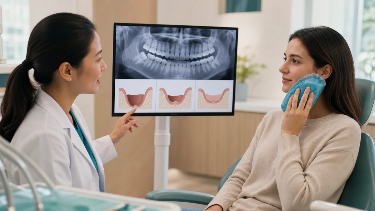
Quick answer: wisdom tooth removal before and after changes are best judged by symptoms and their direction, not by a socket photo alone. Swelling, soreness, jaw stiffness and light oozing can be expected early. Pain should generally become more manageable rather than suddenly worsen. Persistent bleeding, breathing or swallowing difficulty, fever, progressive swelling, or new numbness needs professional advice.
Searching wisdom tooth removal before and after often brings up dramatic photographs. Those images can make a normal recovery look alarming or make a complication look deceptively mild. A photograph cannot show the relationship between roots and a nerve, the condition of the surrounding bone, a sinus connection, the stability of the blood clot, or whether the tooth needed removal in the first place. The safest comparison is a planned clinical assessment before surgery and a symptom-led review afterward.
This guide explains what can reasonably change from the consultation to the first weeks of healing, what usually deserves a routine call, and what needs urgent help. It is educational rather than diagnostic. Your oral surgeon or dentist may give different instructions because the tooth position, surgical difficulty, medical history, medicines and type of anaesthesia differ from person to person. Follow the instructions given by your own clinical team.
1. What wisdom tooth removal before and after pictures can—and cannot—tell you
A useful wisdom tooth removal before and after record may show less facial swelling over several days, improving mouth opening, or a socket that gradually becomes less noticeable. It may also help a clinician understand an external change if you cannot attend immediately. It is not a self-diagnosis tool. Lighting, camera angle, cheek tension and image quality can change how redness, bruising and swelling appear.
Inside the mouth, healing tissue can look pale, cream, yellowish or pink at different stages. Color alone does not confirm infection or dry socket, and scraping or probing the area can disturb healing. A blood clot and early healing tissues are delicate. The important questions are whether pain is improving, whether swelling is stabilizing, whether bleeding is controlled, and whether you feel systemically unwell.
- A photo may show: visible cheek swelling, bruising, limited mouth opening, sutures and the surface appearance of the socket.
- A photo cannot reliably show: nerve proximity, a dry socket diagnosis, deep infection, root fragments, bone healing, sinus involvement or the reason extraction was recommended.
- A safer record: note the day, pain trend, swelling trend, temperature if advised, medicines taken, bleeding, taste or odor, sensation changes and ability to drink.
2. Before removal: confirm why the wisdom tooth is being treated
The “before” stage of wisdom tooth removal before and after begins with diagnosis, not a photograph. A clinician considers symptoms, examination findings and imaging. Common reasons for treatment can include repeated infection around a partially erupted tooth, decay that cannot be restored, damage to an adjacent tooth, gum disease, a cyst or another pathologic change. Pain may have several causes, so the painful area and the radiographic finding need to make sense together.
Not every impacted wisdom tooth must be removed. The UK National Institute for Health and Care Excellence advises against routine prophylactic removal of pathology-free impacted third molars in the NHS and limits surgery to teeth with evidence of disease. The American Dental Association likewise emphasizes examination, X-rays and monitoring of retained wisdom teeth. Recommendations can vary across health systems, and an individual decision should reflect the tooth, the person and the consequences of observation.
Questions to ask before agreeing to extraction
- What specific problem is present, and how certain is the diagnosis?
- What does the panoramic X-ray or other imaging show?
- What are the reasonable alternatives, including monitoring or treating an adjacent problem?
- What are the risks of surgery and the risks of waiting?
- Could a coronectomy be relevant if a lower root is close to a nerve?
- Which anaesthesia or sedation plan is proposed, and what preparation does it require?
- Who should I contact after hours if recovery does not follow the expected course?
3. Imaging changes the meaning of wisdom tooth removal before and after
A panoramic dental X-ray can help show tooth angulation, root shape, adjacent teeth, surrounding bone and the approximate relationship to important anatomy. In selected cases, more detailed imaging may be considered when it could change management. Imaging does not predict the exact appearance of recovery, but it gives the “before” information that a selfie cannot provide.
For lower wisdom teeth, the clinician may discuss the nearby nerve that supplies sensation to the lower lip, chin or tongue. For upper teeth, the maxillary sinus may be relevant. The existence of an anatomical relationship does not mean a complication will occur. It means consent, technique and follow-up should be individualized. A sound wisdom tooth removal before and after plan records baseline sensation before treatment so that a new change can be identified afterward.
In other words, wisdom tooth removal before and after begins with anatomy that only appropriate clinical imaging can reveal.
4. Preparing for wisdom tooth removal before and after care
Preparation depends on whether treatment uses local anaesthetic, sedation or general anaesthesia. Follow the clinic’s exact fasting and medication instructions; do not assume that advice given to another patient applies to you. Tell the clinician about prescription drugs, non-prescription medicines, supplements, allergies, pregnancy, bleeding disorders, sleep apnoea and previous reactions to anaesthesia.
Arrange an adult escort and transport when sedation or general anaesthesia is planned, and follow the team’s restriction on driving, work, machinery and important decisions. Prepare easy-to-eat foods, a clean place to rest, prescribed or approved pain relief, and the clinic’s contact details. The Association of Oral and Maxillofacial Surgeons advises a preoperative consultation, review of imaging and questions about what to expect.
A written wisdom tooth removal before and after plan reduces uncertainty when the anaesthetic wears off and questions arise at home.
A practical before-surgery checklist
- Confirm the appointment, arrival time and fasting window.
- Ask which regular medicines to take or pause; never stop a prescribed medicine without clinical advice.
- Arrange transport and a responsible adult if required.
- Remove uncertainty about payment, consent and after-hours contact before the procedure.
- Stock soft foods and non-alcoholic fluids that fit your medical needs.
- Avoid scheduling demanding work, sport or travel immediately afterward.
- Record any pre-existing numbness, jaw limitation, infection symptoms or facial swelling.
5. Wisdom tooth removal before and after timeline
No single timeline fits everyone. A straightforward erupted tooth and a deeply impacted tooth can produce very different recoveries. The table is a planning guide, not a promise. If your surgeon’s instructions differ, use those instructions.
| Stage | What may be expected | Helpful actions | Reason to contact the team |
|---|---|---|---|
| Before surgery | Examination, imaging, consent, baseline symptoms and anaesthesia planning | Share medical history, medicines and questions; arrange transport if required | New fever, worsening infection, illness, or uncertainty about fasting or medicines |
| Same day | Numbness, soreness as anaesthetic wears off, light blood-stained saliva, fatigue | Bite on gauze as directed, rest, protect the clot, use prescribed or approved pain relief | Bleeding that does not settle with directed pressure, breathing or swallowing difficulty, collapse or severe reaction |
| Days 1–2 | Swelling may build, with bruising, jaw stiffness and tenderness | Soft food, hydration, careful cleaning away from the socket, and the clinic’s cold-pack advice | Rapidly progressive swelling, inability to drink, uncontrolled pain, fever or feeling very unwell |
| Days 3–5 | Swelling should begin to stabilize or improve; soreness may still be present | Continue hygiene and diet progression as instructed; watch the direction of symptoms | Pain that becomes stronger after initial improvement, bad taste or odor with pain, fever, or renewed swelling |
| About one week | Function often improves; the socket remains visible and may trap food | Attend review if arranged; clean only as instructed; do not probe the socket | Persistent severe pain, pus-like discharge, worsening trismus, or concerning sensation change |
| Two weeks and beyond | Soft tissue continues closing while deeper bone remodeling takes longer | Resume activities gradually and maintain routine dental care | Symptoms that fail to improve, new swelling, persistent numbness, or a recurrent opening concern |
6. Wisdom tooth removal before and after in the first 24 hours
The first day is a key point in wisdom tooth removal before and after care. The socket needs a stable clot as part of normal healing. Bite on gauze for the time your team specifies. Light oozing can mix with saliva and look more dramatic than the actual blood volume. Repeatedly checking, spitting, vigorous rinsing, sucking through a straw or touching the socket can disturb the area.
University College London Hospitals advises avoiding rinsing, spitting and touching the socket for the first 24 hours, then beginning cleaning and rinsing only as directed. Rest with your head comfortably elevated if advised. Do not place aspirin or other substances directly on the wound. Use medicines exactly as prescribed or according to the approved label, taking account of allergies, pregnancy, stomach, kidney, liver, bleeding or drug-interaction concerns.
For wisdom tooth removal before and after, protecting the clot is more useful than repeatedly opening the mouth for comparison photos.
Bleeding: expected oozing or urgent problem?
Blood-tinged saliva and a small amount of oozing can occur on the first day. If bleeding restarts, follow the clinic’s pressure instructions with clean gauze and remain seated. If brisk bleeding continues despite correct pressure, fills the mouth repeatedly, causes dizziness, or is accompanied by breathing difficulty, seek urgent care. Never rely on a wisdom tooth removal before and after picture to estimate blood loss.
7. Wisdom tooth removal before and after on days 1 to 3
Swelling commonly becomes more noticeable during the first 48 hours, rather than disappearing immediately after treatment. Bruising may emerge later and change color as it resolves. Jaw muscles can feel tight, making wide opening uncomfortable. These changes are easier to interpret by trend: early swelling that reaches a plateau and then eases is different from swelling that accelerates with fever, spreading redness or difficulty swallowing.
Cold packs are often advised early, but use the timing and skin protection specified by the clinical team. Keep a cloth between a cold pack and skin, and do not sleep with it in place. Some surgeons later advise warmth for jaw stiffness; others do not. Individual instructions matter more than a generic wisdom tooth removal before and after post.
During this phase, a wisdom tooth removal before and after note should emphasize whether swelling is stabilizing and whether fluids remain easy to take.
When swelling needs prompt assessment
Contact the treating team promptly for swelling that keeps increasing after it was expected to settle, fever, pus-like drainage, a bad taste with worsening pain, inability to maintain fluids, or a marked decline in wellbeing. Breathing difficulty, swallowing difficulty, swelling that threatens the airway, severe allergic symptoms or collapse is an emergency.
8. Wisdom tooth removal before and after on days 3 to 5
Dry socket is a painful postoperative complication associated with loss or breakdown of the protective clot. It is not diagnosed simply because a hole is visible. A typical concern is pain that becomes severe or intensifies a few days after extraction, sometimes with an unpleasant taste or odor. Guy’s and St Thomas’ NHS guidance notes that dry-socket pain often appears around days three to five. It needs professional assessment and local care; antibiotics are not automatically the solution.
With uncomplicated healing, soreness may remain but should become more manageable. The socket can still look open, and its surface may not resemble online photographs. If the pain pattern changes sharply, call the clinician rather than inserting cotton buds, rinsing aggressively or applying home remedies. A wisdom tooth removal before and after photo cannot confirm whether the clot is healthy.
The safest wisdom tooth removal before and after comparison here is improving pain versus pain that becomes notably stronger after initial relief.
Dry socket and infection are not the same
Dry socket is mainly a healing disturbance with significant pain. Infection may include worsening swelling, fever, pus-like discharge, increasing redness, a persistent bad taste and feeling unwell, though presentations vary. The two can overlap in symptoms and require examination. Antibiotics should be used only when prescribed for a clinical indication.
9. Wisdom tooth removal before and after in the first week
By the first week, facial swelling and mouth opening often improve, but the socket remains a healing wound. Dissolving sutures may loosen or disappear at different rates. A loose thread is not automatically a problem; do not pull it. If the wound opens widely, bleeding returns, or a suture irritates tissue, ask the clinic whether review is needed.
Food can collect near the area. Clean using the method and timing prescribed by the surgeon. Some patients are given a syringe for later irrigation, while others are not; using one too early or with force can disrupt healing. Do not introduce an unapproved tool because it appeared in a wisdom tooth removal before and after video.
By one week, wisdom tooth removal before and after records should show functional progress even though the socket remains visible.
Why a pale or yellowish surface is not a diagnosis
Early healing tissue may look pale, cream or yellowish in photographs. That appearance alone neither proves pus nor excludes infection. Odor, pain direction, swelling, fever and clinical examination provide more useful context. Do not scrape the surface to see what is underneath.
10. Wisdom tooth removal before and after at week 2 and beyond
Soft tissue gradually narrows the opening, while deeper bone remodeling continues for much longer. The outside can look nearly settled before the socket is fully remodeled. That is why a late wisdom tooth removal before and after image cannot certify complete internal healing. Continue routine oral hygiene and attend a planned follow-up, particularly after a complex extraction, a coronectomy, nerve symptoms or a sinus-related concern.
Occasional mild awareness may occur as chewing returns to normal, but new swelling, renewed strong pain, discharge, fever or deterioration after a quiet period deserves assessment. Persistent altered sensation should also be reported and documented rather than monitored indefinitely without guidance.
A late wisdom tooth removal before and after review is therefore about comfort, function and clinical findings, not just surface closure.
11. Pain relief in wisdom tooth removal before and after care
The goal is manageable pain that trends downward. Use prescribed medicines and written directions. Do not double doses, combine products with the same ingredient, use someone else’s prescription, or assume a medicine is safe because it is available without a prescription. People with ulcers, asthma triggered by anti-inflammatory drugs, kidney or liver disease, anticoagulant use, pregnancy, allergy or other conditions may need tailored advice.
Pain intensity alone is not the only marker. A sudden increase after improvement, pain not controlled by the advised plan, pain with progressive swelling or fever, or pain that prevents drinking should trigger contact. Record timing and medicines taken; that is more useful than repeatedly photographing the socket.
For a meaningful wisdom tooth removal before and after assessment, record the pain trend and exact medicine timing without changing the prescribed plan yourself.
12. Eating and hygiene in wisdom tooth removal before and after care
Choose soft, cool or lukewarm foods initially and chew away from the site when comfortable. Progress texture gradually rather than testing the area with hard, sharp, sticky or seedy foods. Stay hydrated, but avoid very hot drinks while numb and follow anaesthesia-related instructions. A straw can create suction and is often discouraged early.
Clean the rest of the mouth carefully. After the first 24 hours, many clinical instructions introduce gentle rinsing and cautious brushing around the area, but the exact product, frequency and start time should come from the treating team. Vigorous swishing is not better cleaning. If a chlorhexidine product is prescribed, use it for the advised duration and be aware that it can have side effects such as temporary staining or taste alteration.
Good wisdom tooth removal before and after care balances plaque control with protection of the healing socket.
A simple food progression guide
- Early stage: smooth foods that require little chewing, chosen to meet dietary and medical needs.
- As comfort improves: soft eggs, pasta, flaky fish, well-cooked vegetables or similar textures.
- Later: return to normal food gradually, avoiding direct pressure on a tender socket.
- Pause and ask: if swallowing is difficult, nausea prevents fluids, or chewing repeatedly restarts pain or bleeding.
13. Habits and activity in wisdom tooth removal before and after
Smoking is associated with delayed healing and a higher risk of dry socket. UCLH and NHS aftercare guidance advise avoiding it after extraction. Vaping can also involve suction, heat and irritants; ask the surgeon for a realistic cessation plan and support. Avoid alcohol for the period specified, especially when it may interact with medicines or follow sedation.
Rest on the day of treatment. Strenuous exercise can raise blood pressure and restart bleeding, so return gradually according to surgical complexity and clinical advice. Desk work may be possible earlier than physical work, but sedation, sleep disruption, swelling and pain medicine can affect safety. A polished wisdom tooth removal before and after selfie is not evidence that someone is ready to drive or operate machinery.
14. Sensation and sinus checks in wisdom tooth removal before and after
Numbness from local anaesthetic is expected for a limited period. Avoid biting the lip or tongue and take care with hot food until sensation returns. New tingling, persistent numbness, burning or altered sensation in the lip, chin or tongue after lower wisdom tooth surgery should be reported. Early documentation allows the surgeon to assess its distribution and course.
After upper wisdom tooth removal, the surgeon may give sinus precautions if the roots were close to the sinus. Follow instructions about nose blowing, sneezing and pressure changes. Fluid passing between the mouth and nose, unusual air movement at the socket, persistent one-sided sinus symptoms or concern after travel should prompt contact. Do not test the area by forcing air or liquid.
These symptoms show why wisdom tooth removal before and after cannot be reduced to the visible socket alone.
Why baseline sensation matters
Some people already have tingling, pain or altered sensation before treatment. Recording this at consultation separates a pre-existing symptom from a postoperative change. It also improves the usefulness of a wisdom tooth removal before and after clinical review.
15. Coronectomy and wisdom tooth removal before and after
A coronectomy removes the crown while intentionally retaining roots when complete lower wisdom tooth removal may place a nearby nerve at greater risk. UCLH explains that it can reduce nerve injury risk in selected cases, but it is not suitable for every tooth. Infection, root mobility or other findings may change the choice, and retained roots occasionally require later management.
The decision requires imaging, consent and a surgeon’s judgment. A coronectomy should not be presented as universally safer or as a guaranteed way to avoid nerve symptoms. Its wisdom tooth removal before and after pathway also includes follow-up because retained roots can move or rarely become symptomatic.
16. Wisdom tooth removal before and after decision table
| Observation | Usual action | Why |
|---|---|---|
| Mild blood-stained saliva that settles, early swelling, bruising, soreness and jaw stiffness | Follow written aftercare and monitor the trend | These can occur during uncomplicated early healing |
| Pain that becomes stronger around days 3–5, especially after initial improvement | Contact the treating dentist or surgeon promptly | Dry socket or another complication needs examination |
| Worsening swelling, fever, bad taste with pain, pus-like discharge, inability to drink or feeling increasingly unwell | Arrange prompt clinical assessment | Infection, dehydration or another complication may be present |
| New or persistent numbness of lip, chin or tongue; unusual sinus passage symptoms | Report to the surgical team and follow their review plan | Nerve or sinus-related assessment may be needed |
| Brisk bleeding that does not settle with directed pressure, breathing or swallowing difficulty, severe allergic symptoms, collapse or rapidly spreading swelling | Seek emergency help now | Airway, bleeding or severe reaction concerns should not wait for an online reply |
This table supports, but does not replace, the instructions from your clinical team. If you are unsure whether a symptom is urgent, call the treating service or the local urgent-care pathway. In an emergency, use local emergency services.
Use the wisdom tooth removal before and after table to organize a call, not to postpone professional assessment.
17. A 14-point wisdom tooth removal before and after checklist
- Confirm the diagnosis and the reason for removal.
- Review the imaging and nearby nerve or sinus anatomy.
- Discuss observation, complete extraction and any relevant alternatives.
- Share health conditions, medicines, allergies and anaesthesia history.
- Follow individualized fasting and medication instructions.
- Arrange an escort and transport when required.
- Protect the clot during the first 24 hours.
- Track pain and swelling by direction, not by appearance alone.
- Keep hydrated and progress from soft food gradually.
- Clean gently according to the written schedule.
- Avoid smoking and follow advice about vaping and alcohol.
- Return to exercise, work and driving only when safe.
- Report worsening pain, infection signs, bleeding or sensation changes.
- Attend follow-up when planned and maintain regular dental review.
Patients considering treatment with Redent Klinik can read about the clinical team and services on the Redent Klinik English homepage. For a personalized assessment or continuity-of-care question, use the Redent Klinik contact page. Online information cannot determine whether your tooth should be removed.
18. Frequently asked questions about wisdom tooth removal before and after
What should wisdom tooth removal before and after look like on day one?
Day one may include numbness, mild oozing, soreness and early swelling. The socket may be dark because of the clot. Do not keep pulling the cheek, rinsing forcefully or touching the area to inspect it. Follow pressure and medicine instructions, rest and seek help if bleeding does not settle or breathing or swallowing becomes difficult.
Is swelling worse before it gets better?
Swelling often becomes more noticeable during the first 48 hours and then starts to settle. The exact peak varies with the procedure and the individual. A trend of progressive swelling with fever, worsening pain, spreading redness, inability to drink or difficulty swallowing is not something to judge from a photograph; contact a clinician.
How do I know whether the clot is still there?
You usually do not need to inspect or test the clot. The socket’s surface changes during healing and can be difficult to interpret. Protect it by avoiding early vigorous rinsing, spitting, suction and touching. Increasing severe pain a few days later is more important than whether you can see a dark spot.
Does a white or yellow socket mean infection?
Not necessarily. Early healing tissue can appear pale, cream or yellowish. Color alone cannot diagnose infection. Worsening swelling, fever, pus-like discharge, pain that intensifies, persistent bad taste or feeling unwell needs professional assessment. Never scrape the socket.
What does dry socket feel like?
Dry socket commonly causes strong pain that starts or worsens a few days after extraction, sometimes after an initial period of improvement and sometimes with an unpleasant taste or odor. It requires examination and local treatment. A visible hole by itself does not establish the diagnosis.
How long does wisdom tooth removal before and after healing take?
Early pain, swelling and jaw stiffness often improve over days, while soft tissue closure and deeper bone remodeling take longer. Surgical complexity, smoking, health conditions and aftercare affect the course. Use your clinician’s milestones, not a universal deadline or someone else’s photograph.
When can I brush my teeth?
You can usually clean the rest of the mouth carefully, while avoiding trauma to the fresh socket. Many hospital instructions introduce gentle cleaning around the area after 24 hours, but the exact timing and method can differ. Follow the written plan and do not direct a powered brush or high-pressure irrigator into the socket unless advised.
Can I use salt water after wisdom tooth removal?
Some clinical aftercare plans advise gentle warm salt-water rinses beginning after the first 24 hours. The mixture, frequency and duration should follow your surgeon’s instructions. Do not rinse vigorously, and do not start early just because an online post recommends it.
When can I exercise, work or travel?
That depends on the surgery, anaesthesia, bleeding, pain, swelling and job demands. Avoid strenuous exercise early and follow the surgeon’s schedule. Sedation or general anaesthesia creates additional driving and supervision restrictions. Delay travel if it could interrupt urgent follow-up, especially after a complex procedure or a sinus concern.
Is numbness normal after lower wisdom tooth surgery?
Temporary numbness while local anaesthetic is active is expected. New tingling or altered sensation that persists beyond the expected anaesthetic period should be reported to the surgical team. Record the area and whether it changes, but do not pinch or repeatedly test the skin hard enough to cause injury.
Do all impacted wisdom teeth need removal?
No. A symptom-free impacted tooth without pathology may be monitored, depending on the person and applicable clinical guidance. Removal may be recommended when disease or a meaningful risk is identified. NICE discourages routine prophylactic removal of pathology-free impacted wisdom teeth in the NHS. Individual assessment remains essential.
Can wisdom tooth removal before and after photos replace a follow-up?
No. Photos can supplement a conversation, but they cannot assess deep tissues, sensation, bone, clot stability or sinus and nerve relationships. A clinician may still require an examination or imaging. Do not delay urgent assessment while waiting for an online image opinion.
What should I do if I had treatment abroad and develop symptoms at home?
Contact the treating clinic and a local dentist or oral and maxillofacial service. Keep the operation note, imaging, medicine list and contact information. If there is uncontrolled bleeding, airway difficulty, rapidly spreading swelling, collapse or a severe reaction, use local emergency services rather than waiting for international communication.
Medical review: This evidence-based patient guide is prepared for review by Dentist Esma Çevrük Çakır. It prioritizes informed consent, symptom trends, individualized instructions and timely escalation. It does not diagnose a socket, prescribe treatment or replace an in-person dental examination.
19. The safest wisdom tooth removal before and after comparison
The most reliable wisdom tooth removal before and after comparison is not a dramatic image pair. It is a documented reason for treatment, appropriate imaging, informed consent, safe preparation, protected early healing and a clear plan for unexpected symptoms. Normal recovery can look untidy, and a complication can look subtle. Judge the direction of pain, swelling, bleeding, function, sensation and general wellbeing with professional guidance.
If recovery is improving, continue the written aftercare and planned review. If pain suddenly worsens, swelling progresses, fever or bad taste develops, bleeding will not settle, sensation changes, or you cannot drink normally, contact the treating team. Breathing or swallowing difficulty, severe reaction, collapse or uncontrolled bleeding warrants emergency help.
Sources
- NHS: Wisdom tooth removal
- NICE: Guidance on the extraction of wisdom teeth
- NICE: Wisdom teeth removal patient notes
- American Dental Association: Wisdom teeth
- American Dental Association
- AAOMS: Before wisdom teeth surgery
- AAOMS: Potential complications of wisdom teeth extractions
- University College London Hospitals: Dental extraction aftercare
- University College London Hospitals: Coronectomy advice
- Guy’s and St Thomas’ NHS Foundation Trust: Dental surgery and recovery
- MedlinePlus: Tooth extraction
- World Health Organization: Oral health fact sheet