
When you first hear about Invisalign images, you might picture sleek, transparent aligners that gently guide your teeth into their perfect position 😁. But these images are much more than simple visuals—they are windows into real-life dental transformations, patient confidence, and the scientific precision behind one of modern dentistry’s most revolutionary treatments. Whether you’re a potential patient researching your options or a dental professional seeking visual examples to guide consultations, this guide will help you understand why Invisalign images are crucial to both aesthetic outcomes and informed decision-making.
For patients, viewing Invisalign images offers reassurance. It allows them to visualize their potential smile, compare treatment results, and develop realistic expectations. A well-documented gallery of Invisalign before and after photos not only highlights what’s possible but also builds trust. In today’s visual era, where digital impressions and online galleries dominate, these images serve as a powerful communication tool between dentist and patient.
Clinically, Invisalign images play a vital role in diagnostics and progress tracking. Dentists and orthodontists use them at every stage—from initial 3D scanning to final smile review. Modern software allows the creation of time-lapse simulations that demonstrate how teeth will gradually shift. This process ensures precision, consistency, and a clear treatment roadmap. Each image captures small yet essential milestones, helping patients remain motivated throughout their orthodontic journey.
Moreover, Invisalign images demonstrate one of the most unique advantages of aligner therapy: transparency. Unlike traditional metal braces, Invisalign aligners are nearly invisible, which makes before-and-after comparisons even more striking. Many clinics, including Redent Klinik Contact Page, use real patient images (with consent) to highlight these dramatic results. These visuals aren’t just marketing materials—they represent genuine transformations that combine aesthetics and oral health benefits.
But what makes these Invisalign images so impactful is their authenticity. They show the progressive alignment of teeth, the refinement of the bite, and the improvement in facial symmetry. Each picture tells a story—someone’s decision to invest in themselves, to regain confidence, and to smile freely again. Whether the change is subtle or dramatic, these images embody both science and emotion, offering motivation to anyone considering clear aligners.
For dental professionals, Invisalign images serve as diagnostic and marketing tools. By capturing images at specific intervals, practitioners can analyze tooth movement, evaluate patient compliance, and optimize future treatment plans. Many dentists use standardized photographic angles and lighting setups to ensure consistency. They also integrate these visuals into digital platforms and presentations to educate new patients about expected results.
Additionally, the technological foundation behind Invisalign images is evolving rapidly. With AI-powered image analysis, dentists can now simulate treatment outcomes with near-perfect accuracy. Advanced scanning devices create 3D renderings that convert directly into visual progress reports. These developments not only improve efficiency but also enhance patient experience, as individuals can witness their future smiles even before starting treatment.
From a psychological standpoint, the power of Invisalign images cannot be underestimated. Seeing real transformations builds emotional engagement and increases commitment to treatment. Patients who regularly view their progress images are more likely to wear aligners consistently and follow maintenance routines. This visual feedback mechanism strengthens motivation—an essential factor for achieving optimal results in orthodontic care.
For those exploring treatment options, the internet offers vast resources of Invisalign images. However, it’s crucial to distinguish authentic clinical examples from edited or misleading photos. Trustworthy sources include official dental websites, verified orthodontic portfolios, and professional organizations like the American Dental Association. These platforms maintain ethical standards, ensuring that every image reflects genuine, medically supervised transformations.
In summary, Invisalign images bridge the gap between imagination and reality. They inspire confidence, support clinical precision, and demonstrate the life-changing power of modern orthodontics. Whether used to monitor progress, educate patients, or showcase expertise, these visuals are the backbone of transparent and patient-centered dentistry. If you’re curious about how your smile could evolve, take a moment to browse authentic galleries or contact a certified Invisalign provider. Sometimes, one image is all it takes to envision a brighter, more confident you 🌟.
By understanding the meaning and significance of Invisalign images, you’re not just looking at smiles—you’re witnessing technology, artistry, and human transformation in harmony. Every photo tells a success story, and your journey might just be the next one captured in an inspiring before-and-after image.
What Are Invisalign Images and Why Do They Matter?
Invisalign images are detailed photographic or digital records that visually document the process and results of Invisalign treatments. These images go beyond aesthetic appeal—they are essential tools for diagnosis, progress tracking, and patient education. Whether captured by dental professionals or patients themselves, they showcase the gradual transformation of teeth alignment, revealing how Invisalign’s clear aligners achieve natural-looking results over time 🦷.
In orthodontics, visual evidence is as important as clinical data. Invisalign images help both dentists and patients visualize subtle tooth movements that may not be noticeable day-to-day. With each new set of aligners, changes occur incrementally. Regularly photographing these transitions allows for a precise evaluation of how well the treatment is progressing and whether any adjustments are needed. This visual feedback forms the backbone of a transparent and effective orthodontic journey.
Understanding the Clinical Importance of Invisalign Images
From a professional standpoint, Invisalign images are indispensable in treatment planning. Dentists rely on them to verify that the teeth are moving according to the prescribed treatment plan. Digital photos and 3D scans are often compared with the original treatment simulation, ensuring accuracy and patient satisfaction. These images also allow orthodontists to identify areas where the aligners might not be fitting perfectly and make timely refinements to maintain alignment precision.
Another critical function of Invisalign images is their role in communication. In a clinical setting, explaining complex orthodontic concepts to patients can be challenging. However, when patients see their own progress visually, the process becomes much easier to understand. The “seeing is believing” principle helps build confidence and fosters collaboration between doctor and patient. When patients see their teeth shifting in real time through updated Invisalign images, they are more motivated to stay consistent with their aligner wear schedules.
The Psychological Impact of Invisalign Images
Human psychology plays a vital role in treatment success. Regularly viewing Invisalign images can dramatically increase patient satisfaction and motivation. It’s not unusual for patients to feel discouraged during the mid-phase of orthodontic treatment, when results are less obvious. But comparing their current smile to earlier Invisalign images rekindles excitement and boosts morale 💪. Many patients describe this visual progress tracking as a rewarding reminder that every day of disciplined aligner use contributes to their dream smile.
Furthermore, dentists often use these Invisalign images in testimonial galleries to inspire others. When patients witness genuine transformations—especially before-and-after comparisons—they realize that a perfect smile is within reach. These images encourage action and trust, leading more people to choose clear aligners over traditional braces.
How Invisalign Images Improve Patient Education
Patient education is a cornerstone of successful dental care. By incorporating Invisalign images into educational consultations, dentists can better explain expected outcomes and timelines. A visual reference eliminates uncertainty and establishes clear expectations. Patients gain a realistic understanding of how aligners work, how long treatment will take, and what kind of changes to expect after each phase.
For example, during an initial consultation at reputable clinics like Redent Klinik Contact Page, dentists often display sample Invisalign images of similar cases. These real-life visuals help new patients connect emotionally and practically with the process. The images not only illustrate alignment corrections but also improvements in bite functionality, facial harmony, and overall dental aesthetics.
Technological Advances Behind Invisalign Images
Modern orthodontics relies heavily on technology, and Invisalign images have evolved significantly. Earlier, photographs were captured manually with conventional cameras. Today, cutting-edge 3D imaging and AI-driven visualization tools provide hyper-accurate digital simulations of future outcomes. Software such as ClinCheck creates progressive image sequences showing every stage of tooth movement. These predictive visualizations empower both dentist and patient to make data-driven adjustments before treatment even begins.
Additionally, cloud-based platforms allow easy sharing and archiving of Invisalign images. Patients can access their visual progress via mobile apps, while practitioners use the same data to optimize aligner designs. The result is a more transparent, customized, and patient-centered orthodontic experience.
Why Authentic Invisalign Images Build Trust
In an age of social media and online marketing, authenticity is everything. Many individuals seek reassurance through online galleries of Invisalign images, but not all visuals are genuine. Ethical dental practices ensure that every photo shown represents an actual case treated under professional supervision. Organizations such as the American Dental Association emphasize truth in representation, reminding clinics to avoid retouched or misleading imagery.
Authentic Invisalign images reflect real transformations and establish credibility. They show real smiles—not digitally enhanced illusions—helping patients make informed choices. This transparency strengthens brand reputation, fosters patient trust, and reinforces the reliability of Invisalign as a scientifically proven orthodontic solution.
Conclusion: Why Invisalign Images Truly Matter
Ultimately, Invisalign images are more than photographs—they are a visual language of confidence, science, and progress. They help doctors deliver precise, predictable results while empowering patients to stay motivated and informed throughout their treatment. Every snapshot tells a part of a larger success story, from the first aligner tray to the final radiant smile ✨.
When used ethically and strategically, Invisalign images strengthen both clinical outcomes and emotional engagement. They represent the future of orthodontic care—one that’s guided not just by technology, but by visibility, trust, and genuine transformation. Whether you’re a patient starting your journey or a practitioner documenting excellence, these images remind us that every smile has a story worth showing.
Invisalign Before and After Images: Real Transformations
One of the most persuasive ways to understand the true power of Invisalign treatment is through Invisalign before and after images. These visual comparisons illustrate the extraordinary transformations that take place over several months of clear aligner therapy. Seeing real smiles evolve from misaligned to perfectly straight reinforces the effectiveness of the treatment while giving prospective patients confidence in their decision. Whether you’re researching Invisalign for yourself or simply curious about its impact, these Invisalign images reveal the genuine artistry and precision of modern orthodontics 🦷.
The main appeal of Invisalign images lies in their authenticity. They depict the same individual before and after their treatment, often under similar lighting and angles to highlight the exact changes achieved. Unlike promotional models or stock photos, genuine clinical cases display realistic results achieved under professional supervision. For both dentists and patients, these visuals are essential—serving as tangible proof that Invisalign’s clear aligners can deliver both aesthetic and functional improvements.
What Do Invisalign Before and After Images Show?
Each set of Invisalign images tells a story of transformation. The “before” photo typically captures issues such as crowding, gaps, rotated teeth, overbites, or crossbites. The “after” image reveals a beautifully aligned smile that not only looks better but functions more efficiently. These photos often demonstrate improvements in overall facial harmony, as properly aligned teeth enhance jaw balance and lip support.
In some cases, the change is subtle—a slight rotation correction or minor space closure—while in others, it’s a complete realignment of the dental arch. The consistency of results showcased through Invisalign images demonstrates the precision of digital treatment planning and the adaptability of clear aligners for various orthodontic challenges.
The Role of Professional Photography in Invisalign Images
Accurate representation of progress is vital in orthodontic documentation. Professional dental photography ensures that each set of Invisalign images is captured under consistent lighting, exposure, and positioning. Standardized photo angles—frontal, lateral, and occlusal views—are used to document changes objectively. These professional images eliminate distortion and ensure that progress can be analyzed with clinical accuracy.
Moreover, many clinics now use digital scanners and intraoral cameras to generate high-resolution 3D renderings. This digital evolution makes it possible to produce not only still Invisalign images but also animated visualizations that show each stage of movement. As a result, patients gain a complete understanding of how each aligner contributes to the final smile outcome.
Emotional Impact of Invisalign Before and After Images
Beyond science, Invisalign images evoke a strong emotional response. They represent personal victories—people who once hesitated to smile now do so with confidence. These images capture more than teeth; they reflect restored self-esteem and enhanced quality of life 💫. Many patients share their Invisalign journeys online or in clinic testimonials, inspiring others who might feel uncertain about beginning treatment.
Studies in dental psychology show that visual reinforcement increases compliance. Patients who frequently review their own Invisalign images tend to follow instructions more consistently, leading to better and faster outcomes. The emotional satisfaction of seeing real progress strengthens motivation and commitment throughout the process.
Clinical Uses of Before and After Invisalign Images
From a clinical perspective, Invisalign images serve several purposes beyond patient motivation. They are used for documentation, treatment verification, and academic education. Orthodontists review before and after photos to assess treatment accuracy and to refine future plans. For dental education, these images provide valuable visual case studies, allowing practitioners to share outcomes, analyze mistakes, and improve methodologies.
In marketing contexts, authentic Invisalign images also help dental clinics build credibility. Real cases demonstrate transparency and clinical success, reinforcing the trustworthiness of the practice. However, ethical guidelines—emphasized by professional organizations like the American Dental Association—require clinics to use genuine, consent-approved patient photos rather than exaggerated or digitally modified results.
Invisalign Images vs. Traditional Braces Results
When comparing Invisalign images to traditional braces results, one key difference stands out: aesthetics during treatment. Invisalign’s clear aligners make the transformation nearly invisible. Patients can maintain their confidence throughout the process, and the “before and after” comparison looks cleaner and more natural because there are no metal brackets or discolorations caused by wires.
Additionally, Invisalign images often show healthier gums and enamel surfaces after treatment, as removable aligners allow better oral hygiene. This long-term benefit contributes to a more radiant final photo—proof that Invisalign doesn’t just straighten teeth but supports holistic oral wellness.
How to Evaluate Authentic Invisalign Before and After Images
When browsing online galleries or dental websites, it’s important to assess whether the Invisalign images are genuine. Look for signs of consistency—same lighting, same background, identical smile angles. Reputable clinics like Redent Klinik Contact Page maintain strict photographic standards to ensure every image represents a true clinical case. Be wary of overly brightened or digitally retouched photos that distort reality.
Furthermore, consider the diversity of cases displayed. A credible gallery should include various age groups and dental conditions, not just perfect Hollywood smiles. The best Invisalign images show progress across different levels of complexity, proving that clear aligners can help both mild and severe misalignments.
Conclusion: Visual Proof of Modern Orthodontic Excellence
In conclusion, Invisalign before and after images provide undeniable proof of how far dental technology has come. They encapsulate the fusion of digital precision, clinical expertise, and personal confidence. Every smile documented through these Invisalign images stands as a testament to how science and artistry can work together to redefine aesthetics and self-assurance.
For anyone considering treatment, reviewing authentic Invisalign images offers a realistic glimpse of what’s achievable. From subtle alignment corrections to dramatic full-arch transformations, each case demonstrates the reliability and consistency of Invisalign’s clear aligner technology. When guided by certified professionals, these results don’t just improve smiles—they improve lives 🌟.
How to Interpret Invisalign Progress Images Step by Step
Understanding Invisalign images goes far beyond simply looking at before-and-after photos. Each image captured during the treatment journey tells an important story about progress, alignment precision, and biological adaptation. When analyzed correctly, these Invisalign progress images reveal how well a patient is responding to treatment, whether any refinements are needed, and how predictable the final outcome will be 🦷.
Both patients and dentists benefit from knowing how to interpret Invisalign images step by step. Patients gain motivation from visual feedback, while dentists use the data to ensure clinical accuracy. This section explains in detail how these images are captured, evaluated, and used throughout treatment to guide both behavior and adjustments.
Step 1: Establishing the Baseline – The Initial Invisalign Images
Every Invisalign journey starts with a detailed photographic record. These initial Invisalign images include frontal, lateral, and occlusal views taken before the first aligner is worn. They capture not only dental misalignments but also facial proportions, bite alignment, and gum health. These serve as reference points for comparison at every future check-up.
High-quality baseline Invisalign images help orthodontists plan customized treatments using digital simulation tools like ClinCheck. These systems overlay the planned tooth movements onto the patient’s starting images, creating a 3D roadmap of the entire transformation. In other words, your “before” images act as the foundation for your future smile.
Step 2: Monitoring Monthly Progress Through Invisalign Images
Once treatment begins, patients typically switch to a new aligner every one to two weeks. Dentists recommend capturing updated Invisalign images at least once per month to document how each aligner stage contributes to the overall movement. These progress photos are critical for confirming that teeth are tracking according to plan.
Comparing recent Invisalign images to earlier ones helps dentists spot any deviations early. If a tooth isn’t shifting as expected, adjustments can be made—such as adding attachments or revising aligner design. Regular documentation ensures the treatment stays efficient and minimizes the need for costly refinements later.
Professional Tip: Consistency Matters
To accurately interpret Invisalign images, consistency is key. Always use the same lighting conditions, camera angles, and distances. Even minor variations in image perspective can distort the perception of progress. Dental clinics use standardized photographic protocols to ensure each image is comparable to the previous one.
For at-home tracking, patients can follow a simple method: use a neutral background, keep their head straight, and use identical framing for each photo. This makes progress easier to visualize and ensures accurate side-by-side comparisons.
Step 3: Identifying Key Alignment Milestones
During treatment, certain phases represent major progress points. These milestones can be observed directly in Invisalign images. Around week 6–8, you might notice initial tooth rotations correcting. By week 12–16, crowding begins to reduce significantly. Near the end of treatment, bite closure and arch symmetry become apparent.
Professional clinics such as Redent Klinik Contact Page often display anonymized milestone Invisalign images to help patients visualize what to expect at each phase. This visual roadmap motivates adherence to wearing aligners for 20–22 hours a day, a key factor for success.
Reading the Subtle Signs in Invisalign Progress Images
When interpreting Invisalign images, it’s not just about the position of teeth. Look closely at bite alignment, gum symmetry, and midline correction. Even minor differences—such as the spacing between upper and lower arches—indicate significant biomechanical progress. Advanced clinics use digital overlays to measure these changes with millimeter-level precision.
Color and lighting analysis also matter. Healthier gum color in recent Invisalign images often reflects improved hygiene and reduced inflammation, as aligners promote better brushing access compared to braces.
Step 4: Refinement and Final Evaluation
Once the main phase of treatment concludes, dentists conduct a final evaluation using comprehensive Invisalign images. These are compared side-by-side with the initial baseline photos to assess accuracy and patient satisfaction. The final images confirm whether the achieved results match the digital treatment simulation designed at the beginning.
In many cases, dentists create a “before vs. final” composite using professional photo-editing tools—not to alter the images, but to align angles for clearer visualization. This step showcases the precision of Invisalign’s digital orthodontic system and reinforces its credibility as a predictable, high-tech treatment.
AI Integration in Invisalign Image Interpretation
With recent advances in dental technology, artificial intelligence now plays a major role in analyzing Invisalign images. AI software can detect micro-changes in tooth positioning, evaluate patient compliance, and even predict whether refinements are needed before human intervention. These smart analytics not only enhance efficiency but also improve long-term outcomes by catching potential issues early.
Orthodontic AI systems compare thousands of Invisalign images from global databases to identify optimal movement patterns. This ensures personalized care and accelerates progress tracking with unmatched precision.
Step 5: Sharing and Storing Invisalign Images Securely
Privacy and data management are critical when handling Invisalign images. Ethical clinics store photos securely in compliance with health data protection laws. Patients may also access their images through cloud-based apps, allowing them to track their smile transformation anytime 📱.
These visual records serve long-term purposes as well. Should a patient require follow-up care or additional aligners years later, archived Invisalign images provide valuable references for designing new treatment phases.
Conclusion: Mastering the Art of Reading Invisalign Images
Interpreting Invisalign images is both a science and an art. It involves recognizing micro-changes in dental alignment, evaluating bite balance, and understanding patient-specific progress patterns. When done correctly, this process not only ensures clinical success but also deepens patient engagement.
For anyone considering Invisalign, reviewing and understanding these progress visuals can transform the treatment experience. By comparing your personal Invisalign images to clinical benchmarks, you’ll gain insight into how technology and discipline work together to craft your perfect smile. Combined with guidance from trusted professionals and ethical resources like the American Dental Association, this visual awareness empowers you to take full control of your dental journey 🌟.
In short, each snapshot in your Invisalign images collection is more than a photograph—it’s a milestone in your personal transformation story. Learn to read them, and you’ll see your progress with newfound clarity and pride.
Comparing Invisalign Images vs. Traditional Braces Photos
When deciding between clear aligners and metal braces, one of the most convincing comparisons comes from studying Invisalign images side by side with traditional braces photos. These visual contrasts highlight not only aesthetic differences but also variations in comfort, hygiene, and treatment results. The clarity and discreet nature of Invisalign have redefined modern orthodontics, and understanding this transformation through Invisalign images helps patients make more informed decisions 🦷.
Modern dentistry thrives on transparency—both figuratively and literally. The innovation behind clear aligners is visible in every smile captured in Invisalign images. These visuals emphasize natural tooth color, minimal interference, and seamless correction. In contrast, traditional braces photos often display visible wires and brackets, along with minor gum irritation or enamel discoloration. Let’s explore the detailed differences that these images reveal and why Invisalign often emerges as the preferred option.
The Aesthetic Advantage of Invisalign Images
One of the most striking contrasts when analyzing Invisalign images and braces photos is aesthetics. Invisalign aligners are made from medical-grade, transparent SmartTrack material, designed to blend with natural teeth. This allows patients to undergo treatment discreetly without the self-consciousness that metal braces often cause. In before-and-after galleries, Invisalign users typically appear more confident, smiling freely throughout treatment.
Meanwhile, traditional braces photos showcase the mechanical look of brackets and wires. Even though they are effective, they can distract from a person’s natural facial expression. Invisalign images, on the other hand, show smoother transitions and more natural smiles, proving that orthodontic treatment doesn’t have to compromise appearance during the process.
Color and Texture Differences in Dental Photography
In professional dental photography, the difference in color tone and texture between Invisalign images and braces photos is significant. Aligners do not reflect light in the same way metal brackets do, making photographs appear more natural. The absence of metallic glare enhances tooth color accuracy in Invisalign images, allowing for more precise documentation of alignment improvements.
Traditional braces images often show reflections, uneven lighting, and plaque buildup around brackets, which can detract from visual clarity. This aesthetic disparity is one reason why dental clinics increasingly showcase Invisalign images in their marketing and educational materials—they simply photograph better and communicate progress more clearly.
Clinical Insights: Tracking Treatment Progress
Clinically, both Invisalign and braces achieve similar goals—correcting misalignment, closing gaps, and improving bite function. However, Invisalign images provide a clearer view of the progression because there are no obstructing materials on the teeth. Dentists can analyze tooth movement more precisely without visual interference from metal hardware.
Digital scanning systems, such as iTero, automatically generate periodic Invisalign images that track the smallest changes with millimeter-level accuracy. These visuals form a digital timeline of transformation, something that is far more challenging with braces, where brackets obscure the tooth surface. The ability to visually verify treatment success contributes to Invisalign’s reputation for predictability and control.
Ease of Oral Hygiene: A Hidden Factor in the Images
Another subtle difference visible in Invisalign images compared to braces photos is the condition of gum tissue and enamel. Since Invisalign aligners are removable, patients can maintain better brushing and flossing habits. This leads to healthier gums and whiter enamel, which is clearly reflected in mid-treatment Invisalign images.
In contrast, braces often trap food and plaque around brackets, leading to mild discoloration or inflamed gums. These effects are sometimes visible in braces photos and can detract from the overall beauty of the final result. Thus, the clarity and cleanliness seen in Invisalign images are not merely visual advantages—they represent real oral health benefits.
Psychological and Social Impacts Captured in Images
The differences between Invisalign images and traditional braces photos extend beyond appearance—they also reflect emotional well-being. Many Invisalign patients report higher confidence levels during treatment, which shows in their photos. They smile more naturally, attend social events comfortably, and feel less restricted in professional environments 🌟.
Conversely, traditional braces can make patients, especially adults, feel self-conscious. Their smiles in photos often appear more reserved. Invisalign images thus capture not just physical alignment but also psychological transformation—showing the joy and confidence that come from an almost invisible treatment approach.
Time Efficiency and Results in Visual Comparisons
When comparing treatment duration, Invisalign images frequently show comparable or faster results for mild-to-moderate cases. This efficiency is partly due to digital precision and patient compliance. In progress photos, the rate of visible improvement often appears more dramatic in Invisalign images because aligners allow smoother, continuous movement without interruptions from wire adjustments.
Traditional braces still remain ideal for certain complex cases, but visually, the difference in comfort and aesthetics during the process is unmistakable. The ability to smile confidently throughout treatment is a major factor in patient satisfaction, clearly visible in every set of authentic Invisalign images.
Choosing Between Invisalign and Braces: A Visual Decision
Today, patients increasingly rely on visual information to make medical choices. Viewing real Invisalign images alongside braces photos offers clarity about expected results, comfort levels, and lifestyle compatibility. Clinics like Redent Klinik Contact Page often provide side-by-side galleries where patients can assess which treatment fits their aesthetic and practical needs best.
Such transparent visual comparisons empower patients to make confident, evidence-based decisions. When supported by professional counseling and authoritative resources such as the American Dental Association, these images transform curiosity into informed commitment.
Conclusion: Seeing the Future of Orthodontics
In conclusion, Invisalign images offer a cleaner, more transparent, and emotionally uplifting portrayal of orthodontic progress compared to traditional braces photos. They reflect not just aligned teeth but also improved hygiene, confidence, and comfort. The visual harmony captured in these images represents a new standard in orthodontic care—one that prioritizes beauty during the journey, not just at the destination.
For patients seeking aesthetic, predictable, and hygienic treatment, Invisalign images serve as powerful proof of how modern orthodontics blends science with artistry. The clear difference is visible not only in every smile but in the freedom, confidence, and authenticity these images portray ✨.
How Dentists Use Invisalign Images for Treatment Planning
The success of every Invisalign treatment begins with a detailed analysis of Invisalign images. These photographs and digital scans are far more than visual records—they are the foundation of data-driven orthodontic planning. Dentists and orthodontists rely on Invisalign images to diagnose, design, monitor, and refine each stage of a patient’s smile transformation. Without these precise visuals, the accuracy and predictability of the aligner system would not be possible 🦷.
In today’s digital dentistry, Invisalign images function as the bridge between traditional photography and advanced 3D visualization. Each image contributes vital information about tooth position, gum symmetry, jaw alignment, and occlusal relationships. Together, they enable dentists to create individualized treatment plans that ensure every movement is biomechanically efficient and aesthetically pleasing.
The Foundation: Capturing the Initial Invisalign Images
Every Invisalign journey starts with a comprehensive imaging session. Dentists capture high-resolution intraoral and extraoral photographs from standardized angles—front, side, and occlusal. These initial Invisalign images serve as diagnostic references, showing the patient’s natural tooth alignment, bite position, and facial profile before any intervention.
These images are then uploaded into specialized software like ClinCheck, which converts them into 3D models. This process allows orthodontists to simulate tooth movements and visualize the entire treatment from start to finish. The precision of Invisalign images at this stage ensures that even the smallest rotation or spacing adjustment is accounted for, reducing the risk of mid-treatment surprises.
Intraoral Photography and Scanning Technology
Modern practices no longer rely solely on traditional cameras. Instead, advanced intraoral scanners capture digital Invisalign images with exceptional clarity and speed. Devices such as iTero or TRIOS produce accurate digital impressions that feed directly into 3D treatment simulations. This eliminates the discomfort of traditional molds while providing dentists with a more reliable digital record.
Additionally, these scanners can automatically detect changes in enamel texture, gum health, and tooth color—information that traditional photography might miss. The resulting Invisalign images create a full diagnostic picture, ensuring the treatment plan is both functional and cosmetic.
Integrating Invisalign Images into Digital Treatment Planning
Once the baseline Invisalign images are collected, they are integrated into Invisalign’s proprietary software systems. These tools use artificial intelligence and machine learning to analyze the data, predict outcomes, and optimize tooth movement pathways. The dentist can then customize aligner stages according to each patient’s biological response and aesthetic goals.
During this phase, dentists often refer to reference Invisalign images from previous successful cases to predict how similar movements might behave. This comparative analysis enhances planning accuracy and provides patients with realistic expectations through visual previews of their future smiles.
Creating a 3D Roadmap from Invisalign Images
The 3D simulation generated from Invisalign images acts as a digital blueprint for treatment. Each aligner stage is mapped out, showing how individual teeth will shift incrementally. This sequence is reviewed and adjusted by the dentist to ensure biomechanical balance and aesthetic harmony.
In complex cases—such as crossbites or open bites—dentists rely heavily on these images to simulate different corrective approaches before committing to a final plan. This virtual testing saves time, reduces costs, and improves patient satisfaction by avoiding unnecessary trial and error.
Monitoring Treatment Progress Through Invisalign Images
Once treatment begins, dentists use updated Invisalign images at regular intervals to monitor alignment accuracy. Comparing these progress photos with the original simulation allows for precise verification of whether teeth are tracking correctly. If discrepancies appear, refinements can be ordered immediately, ensuring that results remain consistent with the projected plan.
This continuous feedback loop between photography and digital analysis ensures a higher degree of control compared to traditional braces. Each new set of Invisalign images acts as a quality checkpoint, guaranteeing that the patient’s teeth are responding predictably to the aligner sequence.
Refinement Phase: Adjusting Treatment Based on Images
As treatment nears completion, dentists reassess the updated Invisalign images to determine if refinements are necessary. These final adjustments often address small details such as bite closure, tooth angulation, or midline alignment. Without accurate images, detecting these subtle discrepancies would be nearly impossible.
Many clinics, including Redent Klinik Contact Page, emphasize this final imaging phase as crucial to achieving a truly perfect smile. Each refined set of Invisalign images ensures the final result matches both functional and aesthetic goals precisely.
Educational and Communication Value of Invisalign Images
Beyond diagnostics, Invisalign images serve as powerful communication tools. Dentists use them to educate patients about expected progress, explain biomechanics, and maintain engagement throughout treatment. Patients can visually compare their current smile with projected results, making the journey transparent and exciting 🌟.
In clinics that emphasize patient-centered care, these images are often integrated into interactive consultations. Digital displays show side-by-side progress comparisons, helping patients understand the value of compliance and consistent aligner wear. This educational use of Invisalign images improves patient trust and motivation.
Professional Collaboration and Case Sharing
Invisalign’s cloud-based platforms allow secure sharing of Invisalign images between dental professionals worldwide. This feature enables multi-specialist collaboration—for instance, between orthodontists, prosthodontists, and cosmetic dentists. Experts can analyze complex cases collectively, leveraging different perspectives to optimize treatment outcomes.
Additionally, these shared Invisalign images serve as learning material in academic dentistry. Universities and continuing education programs use them to train new dentists on interpreting visual data and planning precise orthodontic movements. Such visual learning enhances the accuracy and predictability of future treatments.
Conclusion: Precision Dentistry Through Visual Intelligence
In essence, Invisalign images are the backbone of modern orthodontic treatment planning. They enable dentists to visualize, design, and refine each stage of movement with unparalleled precision. From the first photo to the final smile, these visuals combine artistry and technology to produce predictable, life-changing results.
When used ethically and accurately—supported by reputable sources like the American Dental Association—Invisalign images elevate the entire treatment process. They transform abstract dental data into tangible visual progress, helping patients see and understand their transformation before it’s even complete. This visual intelligence defines the future of dentistry, where every smile is designed with science, verified by imagery, and perfected through patient collaboration 💫.
The Best Places to Find Authentic Invisalign Images Online
In the digital age, patients often turn to the internet to research treatments before visiting a dental clinic. When it comes to Invisalign, one of the most helpful resources is authentic Invisalign images—real photos of people who have undergone clear aligner treatment. These images provide visual proof of results, helping potential patients build realistic expectations and confidence. However, not all visuals online are trustworthy. Knowing where to find genuine, medically accurate Invisalign images can make all the difference between informed decision-making and misleading impressions 🦷.
This guide explores where to find the most reliable and verifiable Invisalign images online, how to identify authenticity, and which professional organizations regulate visual content standards in dentistry. Whether you’re a patient seeking inspiration or a dental professional curating educational material, the sources below ensure credibility and quality.
1. Official Invisalign Website and My Invisalign App
The most direct and authentic source of Invisalign images is the official Invisalign website. It features a dedicated section for “Before and After” cases submitted by certified providers around the world. Each image displayed on the site has been verified by Invisalign and adheres to strict photographic standards. The platform includes filters by treatment type—crowding, gaps, bite correction—and displays detailed case durations.
The My Invisalign App, available on iOS and Android, also allows users to upload their own Invisalign images for tracking progress. These uploads are often anonymized and added to the community gallery, creating an expansive library of authentic real-world cases. Users can even view stories from others who share similar alignment challenges, making the visual experience both personal and educational.
Why the Official Invisalign Sources Are Reliable
These platforms employ ethical review systems that ensure all Invisalign images are genuine, unaltered, and taken under consistent photographic conditions. No filters or editing tools are used, which guarantees that the results reflect true clinical progress. Patients can trust these visuals to represent medically accurate outcomes rather than marketing exaggerations.
2. Dental Clinic Websites and Professional Portfolios
Many certified dental clinics maintain galleries showcasing their real patients’ Invisalign images. For example, reputable institutions like Redent Klinik Contact Page offer transparent, case-by-case visuals showing transformations over time. These galleries often include full case descriptions, such as duration, number of aligners used, and complexity level.
When browsing clinic-based Invisalign images, look for transparency indicators—patient consent statements, dentist names, and verifiable clinic credentials. Ethical practices clearly state when an image has been shared with permission, often accompanied by testimonial videos or clinical notes. This ensures authenticity and patient privacy are maintained simultaneously.
How to Identify Authentic Clinic-Based Images
Authentic Invisalign images from clinics typically have consistent lighting, identical framing between “before” and “after” photos, and minimal retouching. Overly brightened or airbrushed photos can be a red flag. Real cases may show slight differences in lip position or skin tone—these variations are signs of genuine photography rather than digitally manipulated advertising.
3. Reputable Dental Associations and Educational Platforms
Another excellent source of verified Invisalign images comes from professional dental associations. Organizations like the American Dental Association and the British Orthodontic Society publish educational galleries and research materials featuring real Invisalign cases. These resources are particularly valuable for dental students, educators, and clinicians who require visual case studies for academic analysis.
In many cases, these associations host case libraries within their online journals or continuing education portals. The Invisalign images here are documented with technical details—case duration, dental arch form, torque correction, and material response. Such accuracy transforms these photos into scientific evidence rather than promotional material.
Why Educational Sources Matter
Educational repositories promote transparency and uphold professional integrity. They showcase the diversity of orthodontic cases—from minor cosmetic improvements to full bite corrections—ensuring that users understand the wide applicability of Invisalign technology. Moreover, these Invisalign images help counter misinformation by providing standardized, peer-reviewed visuals from recognized experts.
4. Patient Communities and Social Media Platforms
While social media should be approached cautiously, it can still be a valuable source of real Invisalign images—especially from verified patient accounts. Platforms like Instagram, Reddit, and Facebook host communities where users document their Invisalign journeys week by week. These personal logs offer authentic insights into the emotional and physical aspects of treatment 🌟.
However, users should be careful to differentiate between verified and promotional content. Authentic posts often include captions detailing treatment stages, aligner counts, or clinic names, while overly polished or model-like images may be part of influencer marketing campaigns. Engaging with real patient stories can be inspiring, but professional guidance should always accompany online research.
Tips for Evaluating Social Media Invisalign Images
- Check for consistent lighting and camera angles across progress posts.
- Read captions carefully—genuine users often share challenges, discomfort experiences, or tips.
- Look for mentions of certified clinics or orthodontists who supervised the case.
- Be cautious of results that seem “too perfect” or lack any timeline information.
5. Academic Case Studies and Dental Journals
For those seeking in-depth, technical perspectives, academic journals provide highly reliable Invisalign images. Publications like the Journal of Clinical Orthodontics and the American Journal of Orthodontics and Dentofacial Orthopedics frequently include clinical case studies with photographic documentation. These images undergo strict peer review to confirm their accuracy and scientific value.
In such publications, Invisalign images are used not only to showcase outcomes but also to illustrate complex biomechanical principles—such as anchorage control, torque management, and sequential tooth movement. This level of transparency ensures that professionals and students alike can learn from real data-driven visual examples.
Conclusion: Trust Authenticity, Not Aesthetics
Finding genuine Invisalign images online requires discernment, but the right sources can empower patients to make confident choices. Official Invisalign platforms, reputable dental clinics, and professional associations provide the most trustworthy visuals, ensuring that every image represents real progress and scientifically validated results.
By relying on verified Invisalign images instead of edited marketing content, patients gain accurate expectations about treatment length, comfort, and outcomes. As dental transparency continues to evolve, these authentic visuals stand as proof that science—not digital enhancement—is what truly creates the perfect smile 😁.
How to Take Perfect Invisalign Images at Home 📸
Capturing clear and accurate Invisalign images at home is one of the most effective ways to monitor your orthodontic progress between dental visits. These self-taken images help both you and your dentist visualize tooth movement, track treatment milestones, and maintain motivation throughout your journey. When done correctly, at-home Invisalign images can mirror the quality of professional photographs and serve as reliable documentation for remote consultations 🦷.
However, not all at-home photos are created equal. Blurry, poorly lit, or inconsistent shots can make it difficult to assess progress accurately. That’s why understanding the correct technique, lighting setup, and positioning is essential. In this section, you’ll learn step-by-step how to capture professional-quality Invisalign images at home using basic tools and proper guidance.
1. Preparing for Your At-Home Invisalign Photos
Before you start taking Invisalign images, ensure that your environment is clean, well-lit, and distraction-free. Natural light works best—try facing a window or standing in a brightly illuminated room. Avoid overhead lights that cast shadows on your face. If natural light isn’t available, use a ring light or two lamps placed at 45° angles to evenly illuminate your smile.
Next, prepare your equipment. You don’t need a professional camera; most modern smartphones can produce high-quality Invisalign images when used correctly. Wipe your camera lens clean, switch to portrait mode, and use a timer or tripod to minimize shaking. Have a mirror ready for side shots, and if possible, use dental retractors (or two clean spoons) to hold your lips away from your teeth for a full view of your alignment.
Checklist Before Capturing Invisalign Images
- Ensure your aligners are either removed or cleaned thoroughly if photographing with them on.
- Rinse your mouth and brush your teeth for a clear image without residue.
- Use consistent backgrounds and lighting for every session.
- Keep your camera at mouth level for all Invisalign images.
2. Capturing Front, Side, and Occlusal Views
To create a complete photographic record, capture three main perspectives in your Invisalign images: front, side, and occlusal (top and bottom). Each angle offers unique diagnostic information.
Front View: Smile naturally while showing your teeth fully. The camera should be centered at the midline of your face, capturing both upper and lower arches evenly. This view allows you and your dentist to assess symmetry, arch shape, and bite closure.
Side View: Turn your head slightly (about 45 degrees) and photograph both the left and right sides. These Invisalign images help evaluate canine positioning and molar occlusion, showing how well the upper and lower teeth fit together.
Occlusal View: These are the “bird’s-eye” shots of your upper and lower arches. Tilt your chin upward to capture the upper arch and downward for the lower arch. Use a mirror if necessary to maintain focus. These images are critical for identifying crowding or spacing issues.
Technical Tips for Sharp Invisalign Images
To avoid blur, hold your breath while taking the shot, use a timer delay, and keep your arms steady. Activate your phone’s gridlines to center your teeth and maintain consistent framing. Always take multiple Invisalign images from each angle and keep the clearest ones for comparison.
3. Using Apps and Tools to Improve Image Quality
Several smartphone apps can assist in capturing and organizing Invisalign images. Apps like the “My Invisalign” tracker or “DentalMonitoring” allow you to upload your photos, compare them to earlier stages, and even share them securely with your dentist. Some apps provide alignment overlays that help ensure you capture images from the same angle each time.
These technologies make it easier to track subtle progress between appointments, ensuring your teeth are moving according to plan. Regularly reviewing your Invisalign images also helps you notice small improvements, keeping you motivated to wear your aligners consistently.
Lighting and Background Optimization
For professional-looking Invisalign images, use a neutral background—plain white or light gray works best. Avoid colorful or patterned walls that can distract from your teeth. If using artificial lighting, aim for soft, diffused brightness. Avoid harsh shadows or reflections caused by flash photography, as they can obscure details like enamel texture or aligner fit.
4. Consistency Over Time: Building a Progress Library
One of the greatest benefits of at-home Invisalign images is tracking progress over time. Schedule a specific day and time every week (for example, every Sunday morning) to take your photos. Consistent timing ensures that lighting, angle, and exposure remain similar, making comparisons easier and more accurate.
Label each folder with the date and aligner number, such as “Week 6 – Aligner #3.” This organization method creates a timeline that can be shared with your orthodontist during online consultations or in-person checkups. Over several months, you’ll build a visual story of your journey—something many patients find encouraging and rewarding 🌟.
Data Privacy and Sharing Considerations
When sharing Invisalign images online or via apps, prioritize privacy. Use secure, HIPAA-compliant platforms recommended by your dentist. Avoid uploading your personal progress images to public social media unless you’re comfortable sharing them. Clinics like Redent Klinik Contact Page provide secure upload systems to keep patient data confidential while maintaining professional communication.
5. When to Contact Your Dentist
While self-monitoring with Invisalign images is empowering, it’s important to recognize when professional input is needed. If your progress photos show unexpected gaps, misalignments, or changes in gum health, contact your dentist promptly. Dentists use your submitted Invisalign images to evaluate aligner fit and tooth movement accuracy remotely. They can determine whether an in-person adjustment or refinement set is necessary.
In some cases, Invisalign images reveal early signs of non-compliance or incorrect aligner wear. Addressing these issues quickly prevents delays and ensures that your final results match the treatment plan exactly.
Conclusion: Empowering Your Smile Through At-Home Invisalign Photography
Learning to capture clear and consistent Invisalign images at home enhances communication with your dentist, boosts motivation, and adds transparency to your treatment process. With just a smartphone, good lighting, and a few minutes of preparation, you can produce professional-quality images that document your transformation in detail.
By following these guidelines—and cross-referencing your progress with trusted professional advice or resources like the American Dental Association—you’ll gain greater control and confidence in your Invisalign journey. Every photo you take becomes part of your smile’s evolution, showing that the path to perfection can be both visual and empowering 📸✨.
Understanding Invisalign Smile Simulation Images
Before beginning orthodontic treatment, many patients want to know exactly what their future smile will look like. This is where Invisalign images—specifically smile simulations—play a crucial role. These digitally generated visuals help patients visualize projected results, track real-time changes, and feel confident in their decision. Smile simulation technology transforms complex dental data into a clear, lifelike preview of post-treatment outcomes 🦷.
Unlike standard before-and-after photos, Invisalign images in simulation form are created using artificial intelligence and 3D modeling software. They merge digital scans, bite alignment data, and dental movement algorithms to create precise, predictive visuals. By analyzing these simulations, both patients and clinicians can understand the treatment’s expected progress and outcome long before the first aligner is worn.
1. What Are Invisalign Smile Simulation Images?
Invisalign simulation images are advanced digital renderings that show predicted tooth movement over time. Generated by software such as ClinCheck, these images represent each aligner stage in sequence—from the initial misalignment to the final straightened result. Essentially, they serve as a virtual “roadmap” of your smile transformation.
For patients, viewing these Invisalign images eliminates uncertainty. They can see their future smile from multiple angles—front, side, and occlusal—helping them visualize how their teeth and facial symmetry will change. For dentists, simulations act as planning blueprints, allowing precise control over tooth movement, anchorage, and timing.
How Simulation Differs from Real Invisalign Images
Traditional Invisalign images show real photographic results, whereas simulations are digital predictions. Although both serve visual purposes, simulations provide forward-looking insight, while photographic documentation confirms actual progress. When combined, they form a complete visual journey from projection to reality.
Moreover, simulation-based Invisalign images can be updated throughout treatment. If aligners are not tracking correctly or adjustments are made, the simulation adapts automatically, offering patients the reassurance that their treatment remains on course.
2. The Technology Behind Invisalign Smile Simulations
Creating Invisalign images for smile simulations involves cutting-edge digital dentistry tools. It starts with a 3D intraoral scan using a device like iTero. This scanner captures thousands of images per second, converting them into a high-resolution 3D model of your teeth and gums. The model is then analyzed by Invisalign’s AI algorithms, which calculate optimal tooth movements across the entire treatment plan.
Each step—every micro-movement—is visualized through a progression of Invisalign images within the software. These stages can be viewed interactively, allowing the dentist to rotate, zoom, and assess the plan from all angles. The result is a highly detailed, medically precise visual forecast of your smile transformation.
AI and Machine Learning in Simulation Accuracy
Artificial intelligence enhances the reliability of Invisalign images by comparing your scan data to millions of successful cases worldwide. This vast database allows the software to predict biological responses and movement timing with impressive accuracy. The more data it processes, the smarter it becomes—improving both clinical precision and patient confidence.
AI-driven Invisalign images also help dentists predict potential complications early, such as teeth that may resist movement. These insights make treatment safer, faster, and more comfortable for patients.
3. Benefits of Smile Simulation for Patients
Seeing a realistic preview of your future smile is both motivating and reassuring. Invisalign images in simulation form allow patients to make informed decisions about their orthodontic investment. By viewing their predicted results, patients understand the process, duration, and benefits of treatment more clearly.
Clinics that offer simulations, such as Redent Klinik Contact Page, often find that patient engagement and satisfaction increase significantly. Patients become active participants in their own treatment, regularly comparing their live progress photos to the digital simulations. This involvement encourages consistent aligner wear and boosts overall compliance 🌟.
Emotional and Psychological Advantages
Beyond clinical accuracy, simulation-based Invisalign images provide emotional reassurance. Many patients feel nervous about committing to orthodontic care, especially adults concerned with appearance during treatment. Seeing their future smile helps them envision not just straighter teeth, but renewed confidence, better speech articulation, and improved facial balance.
Psychologically, these visuals serve as motivational anchors. When patients encounter plateaus or doubt their progress, revisiting their original Invisalign images reminds them of how far they’ve come—and how close they are to their dream smile.
4. Dentist and Patient Collaboration Through Simulation
Another remarkable aspect of simulation technology is its ability to foster collaboration. Dentists can adjust digital Invisalign images interactively—rotating teeth, widening arches, or refining bite alignment—and instantly show patients the difference. This visual dialogue enhances transparency and builds trust, as patients see their feedback directly influencing the plan.
Furthermore, these Invisalign images can be shared remotely via secure cloud systems. Patients traveling or living abroad can maintain consistent communication with their dental team, reviewing updated simulations and receiving virtual checkups. This accessibility is transforming how orthodontic care is delivered worldwide.
Clinical Precision Through Data Visualization
Each simulated image includes precise metrics—angular rotation, torque levels, and predicted movement velocity. Dentists can adjust these variables to ensure optimal balance between speed and biological safety. This integration of data science and visual imaging turns Invisalign images into powerful diagnostic tools that improve long-term treatment stability.
5. Evaluating the Accuracy of Invisalign Simulation Images
Many patients wonder how closely the simulated Invisalign images match the final results. Studies show that modern Invisalign simulations are over 90% accurate in predicting tooth positioning when aligners are worn as prescribed. The small discrepancies that do occur typically result from inconsistent aligner wear or unexpected biological variations.
To ensure accuracy, dentists frequently update Invisalign images throughout the treatment process. By aligning real progress photos with simulation stages, they confirm whether adjustments are required. This continuous verification process guarantees that the digital prediction and clinical reality remain in sync.
Conclusion: The Future of Orthodontics Through Digital Visualization
In today’s era of precision dentistry, Invisalign images in the form of smile simulations represent the perfect fusion of technology, artistry, and science. They transform invisible aligners into visible inspiration, allowing patients to preview the future and witness each stage of their journey with confidence. From AI-powered accuracy to interactive 3D visualization, these images are revolutionizing how both patients and professionals approach orthodontic care.
Trusted organizations like the American Dental Association continue to endorse transparent, evidence-based imaging standards, ensuring that every simulation remains a reliable reflection of real results. For patients, these Invisalign images are more than digital renderings—they’re a glimpse into the future of their own smile, where science and self-confidence meet in perfect harmony ✨.
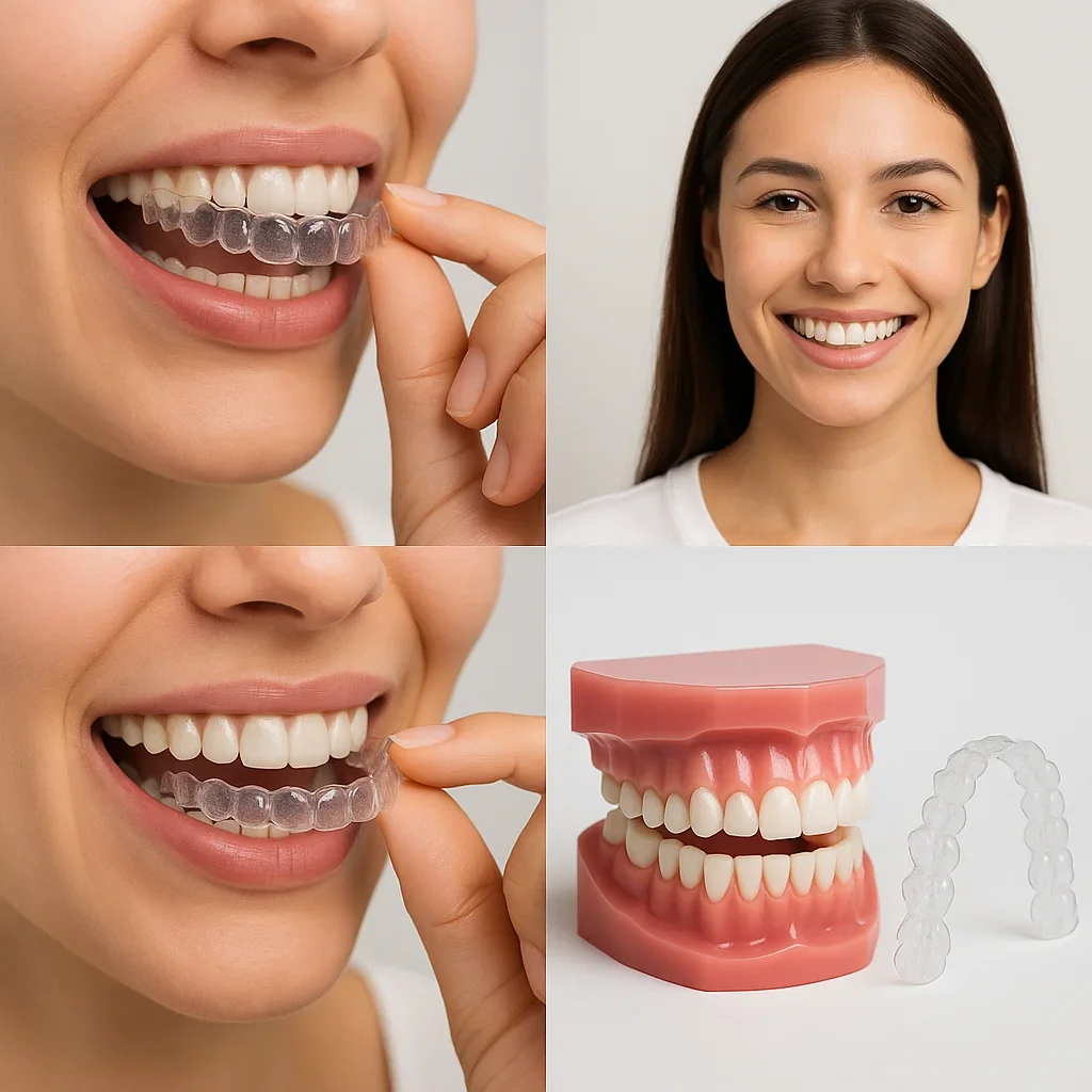
How Invisalign Images Help Track Progress and Ensure Results
One of the most powerful features of modern orthodontic care is the ability to visualize progress in real time through Invisalign images. These photos, taken periodically throughout treatment, are not just for aesthetics—they are essential diagnostic tools that enable dentists to ensure accuracy, identify potential deviations, and maintain treatment efficiency. For patients, seeing measurable progress through visual documentation boosts motivation, reinforces compliance, and builds confidence in their journey toward a perfect smile 😁.
When used consistently, Invisalign images allow both patients and practitioners to collaborate effectively. They bridge the gap between digital treatment planning and real-world results, creating a transparent and data-driven orthodontic process. This section explores how progress tracking with Invisalign photos ensures precision, enhances communication, and secures long-term success.
1. Why Tracking Progress with Invisalign Images Matters
Every smile transformation with clear aligners is a carefully orchestrated process. Invisalign images provide the visual feedback necessary to confirm that teeth are moving as planned. Without them, even small deviations could go unnoticed, potentially delaying results or compromising alignment.
For dentists, periodic photo reviews are like clinical checkpoints—they verify that tooth movements correspond with digital simulations. For patients, reviewing their own Invisalign images helps them appreciate the progress made and reinforces consistent aligner wear. These regular photographic comparisons also assist in detecting issues early, such as incomplete seating, minor rotation errors, or improper aligner fit.
From Data to Visual Precision
While software provides numeric tracking data, visual evidence from Invisalign images adds a critical layer of clarity. For example, subtle improvements in symmetry or midline correction are often easier to see than to measure. By combining digital metrics with photographic progression, dentists gain a complete picture of the treatment’s accuracy and effectiveness.
2. Establishing a Progress Schedule
Consistency is key to meaningful comparison. Most orthodontists recommend capturing Invisalign images every 2–4 weeks or at each aligner change. These intervals allow enough time for visible differences while keeping deviations manageable. The first image acts as a baseline, while subsequent ones form a timeline that visually narrates the treatment journey.
Many clinics, including Redent Klinik Contact Page, follow a structured protocol for documenting each phase of treatment. By maintaining consistent lighting, angle, and framing, they ensure the Invisalign images remain scientifically comparable. The resulting sequence allows both dentist and patient to assess transformation speed and stability over time.
Recommended Angles for Progress Documentation
- Frontal View: Displays overall tooth alignment and bite evolution.
- Left and Right Lateral Views: Capture molar relationships and jaw coordination.
- Occlusal Views: Reveal changes in arch shape and spacing between teeth.
- Smile View: Highlights aesthetic improvement and facial harmony.
3. Comparing Progress with ClinCheck Simulations
One of the biggest advantages of Invisalign treatment is its integration of digital planning. By comparing periodic Invisalign images with the ClinCheck simulation, dentists can instantly verify whether real progress aligns with projected outcomes. If discrepancies arise, refinements can be made early—avoiding the need for lengthy mid-course corrections later.
This comparison is particularly useful in complex cases involving bite correction, rotations, or vertical movements. Real-world Invisalign images help confirm that every aligner stage is being completed as predicted. When deviations are detected, additional aligners (refinement trays) are designed to recalibrate progress, ensuring precise final results.
AI-Powered Image Matching
Advanced AI software now allows automatic alignment between real and simulated Invisalign images. These tools overlay photos onto digital models, highlighting differences in tooth position, rotation, or arch width. Such technology saves time, increases diagnostic accuracy, and enables remote monitoring of patients who can upload their progress photos securely through tele-dentistry platforms.
4. Enhancing Patient Motivation and Compliance
Tracking progress through Invisalign images isn’t just a technical process—it’s also psychological. Many patients underestimate how much their smile has improved until they compare photos side by side. Seeing visible progress can reignite enthusiasm, especially during the slower mid-phase of treatment when motivation tends to dip 🌟.
Clinics that share milestone Invisalign images with patients report higher adherence rates and fewer treatment delays. Some even create progress galleries where patients can view their evolution month by month, reinforcing the reward of consistent aligner wear. These visual affirmations strengthen accountability and turn orthodontic care into an empowering, collaborative experience.
Patient Engagement Tools
Modern platforms and apps integrate progress tracking with Invisalign images. For instance, the My Invisalign App allows users to upload photos, compare them to initial stages, and receive automated feedback. Such features transform patient engagement, making treatment feel interactive rather than mechanical. The sense of ownership these visuals provide significantly enhances satisfaction and treatment success.
5. Professional Review and Long-Term Stability
Once treatment nears completion, dentists conduct a final analysis using all accumulated Invisalign images. Comparing the first and last images validates the alignment’s success and ensures that retention plans are appropriately designed. These records also serve as a visual reference for future dental work, such as cosmetic enhancements or bite maintenance.
Furthermore, long-term follow-ups at 6 and 12 months post-treatment often include updated Invisalign images. These confirm that teeth remain stable and that retainers are functioning as intended. For both dentist and patient, this ongoing documentation provides peace of mind and professional accountability.
6. Ethical and Educational Use of Invisalign Images
High-quality, consented Invisalign images often serve educational purposes in dental training and research. By analyzing thousands of documented cases, orthodontists identify patterns that improve future treatment protocols. Organizations like the American Dental Association emphasize ethical use of patient visuals, ensuring they are shared only with explicit consent and for professional advancement.
Clinics that maintain detailed photo libraries also contribute to public awareness. When shared responsibly on official platforms, Invisalign images inspire others considering aligner therapy, showing real transformations achieved through science-backed orthodontic care.
Conclusion: A Picture-Perfect Journey Toward a Confident Smile
Invisalign has revolutionized orthodontics, but it’s the power of Invisalign images that truly brings the transformation to life. From initial documentation to final review, these visuals ensure that every movement is precise, every milestone celebrated, and every result predictable. By combining clinical accuracy with visual motivation, Invisalign creates a transparent partnership between dentist and patient—one where progress is visible, measurable, and deeply rewarding.
Whether for personal satisfaction or professional validation, the consistent use of Invisalign images represents the gold standard in modern dental monitoring. With every photo, patients witness not only the evolution of their smile but the tangible results of dedication, technology, and trust in expert care 💫.