
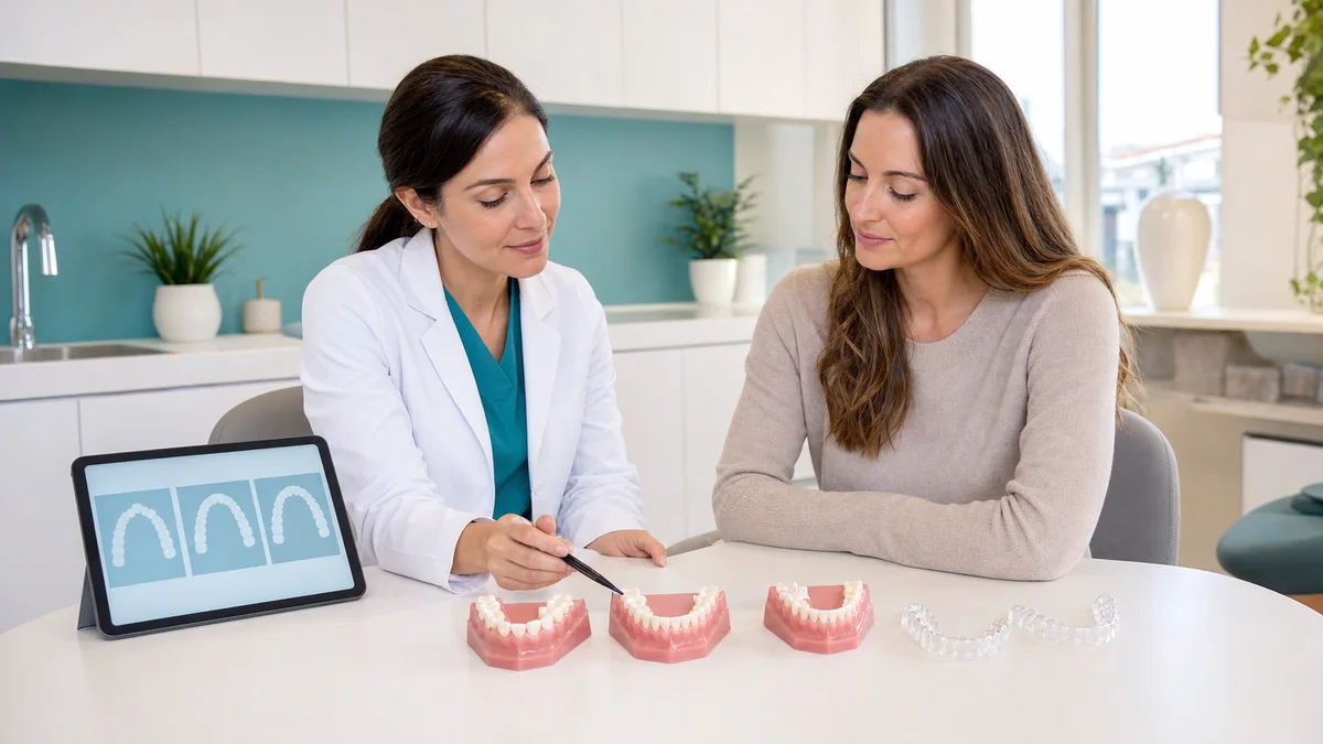
Searching invisalign before after crowding often starts with a simple visual question: can clear aligners make overlapping teeth look straighter? They can be an option for selected crowded dentitions, but a photograph shows only part of an orthodontic result. It may reveal crown alignment from one angle while hiding root position, gum support, bite contacts, arch width, tooth wear and whether the result remained stable after active treatment.
Invisalign® is a commercial clear aligner brand. Clear aligners are a broader category of sequential removable orthodontic appliances. The American Association of Orthodontists explains that aligners can treat some crowded teeth, while also emphasizing that they are not suitable for everyone and that an orthodontic professional should decide whether aligners, braces or a combined approach best fits the diagnosis. A brand name does not replace that individualized decision.
This guide is designed to help patients read before-and-after material without turning it into a promise. It explains what a fair comparison needs, which clinical records sit behind the photographs, how space may be created, why refinements and retainers matter, and which questions to ask before consent. It does not diagnose crowding, rank a personal case from a selfie or predict a treatment result.
1. What Dental Crowding Actually Means
Crowding means that teeth do not have enough suitable space to occupy healthy, planned positions in the dental arch. Teeth may overlap, rotate, erupt inside or outside the arch, or lean in ways that make cleaning and bite coordination more difficult. The visible amount of overlap is not a complete measure of severity because the roots and supporting bone may tell a different story.
Crowding can involve one area or both arches. It may be related to tooth size, arch dimensions, eruption pattern, early tooth loss, retained teeth, missing teeth, previous orthodontic relapse or a combination of factors. A clinician also considers the relationship between upper and lower jaws. Simply straightening the front crowns without planning the bite can exchange one visible problem for another.
The AAO describes clear aligners as custom trays that apply sequential pressure and identifies crowded teeth among conditions they may treat. Suitability still depends on the movements needed. Large rotations, root control, severely displaced teeth, impacted teeth, skeletal discrepancies or unstable periodontal support may require auxiliaries, fixed appliances, another discipline or a different strategy.
That is why an invisalign before after crowding comparison should begin with the original diagnosis rather than the attractiveness of the final smile. If the starting problem is not defined, the viewer cannot know what the images demonstrate.
2. Invisalign Before After Crowding: A 10-Point Image Audit
Use the table below when a clinic, manufacturer, influencer or patient presents a transformation. It does not decide whether the outcome is good; it identifies information needed for a responsible interpretation.
| Comparison check | What a fair record should show | What can mislead | Question to ask |
|---|---|---|---|
| 1. Same patient | Clear confirmation that both records belong to one consented case | Unrelated images or unclear provenance | Is this a documented clinical series? |
| 2. Same view | Matching front, side and biting-surface angles | One unfavorable “before” angle and one flattering “after” angle | Are the camera and head positions comparable? |
| 3. Same expression | Similar lip posture and smile effort | A relaxed before image compared with a broad after smile | Can I see retracted intraoral views as well? |
| 4. Similar lighting | Consistent exposure, white balance and magnification | Whiter, brighter or retouched final teeth | Were color and contrast altered? |
| 5. Both arches | Upper and lower occlusal views | Only the most improved arch is shown | How did the other arch change? |
| 6. Bite together | Front and side records with teeth in a reproducible bite | Separated teeth or a posed jaw hiding contacts | Was the bite clinically assessed? |
| 7. Gum margins | Sharp enough images to inspect recession, swelling and black triangles | Cropped or blurred gum edges | How did periodontal findings change? |
| 8. Timing | Date or phase identified: active finish, refinement or retention | An “after” photo taken before stability is assessed | Was this taken after retainer delivery? |
| 9. Additional treatment | Disclosure of IPR, attachments, elastics, extraction, bonding or whitening | Attributing every visual change to trays alone | What else occurred between the images? |
| 10. Clinical context | Diagnosis, goals and limitations explained without identifying the patient | A result image used as a universal guarantee | Why is this case relevant to mine? |
Even a well-standardized series is evidence about one individual, not a forecast for another. Anatomy, periodontal support, cooperation, movement complexity and treatment goals differ. A gallery can support a conversation; it should not replace a personal assessment.
3. Standardized Photographs Make Comparisons More Honest
Orthodontic photographs are clinical records when they are taken consistently. Common views include the face at rest and smiling, retracted front and side bite views, and occlusal views of the upper and lower arches. The exact record set can vary, but matching views allow a clinician to examine changes rather than changes in presentation.
Small photographic differences can exaggerate an apparent transformation. A closer camera position makes front teeth seem larger. Head rotation changes midline appearance. A higher camera angle can hide lower crowding. Stronger smile effort displays more teeth. Bright exposure can make enamel look whiter, while dehydration during a clinical photo can temporarily change tooth appearance.
Patients should also distinguish alignment from cosmetic editing or other dentistry. Whitening, edge bonding, veneers, gum treatment or professional cleaning can alter the final appearance without being caused by orthodontic movement. Those services may be entirely appropriate, but they should be disclosed when images are presented as an aligner outcome.
- Look for equivalent front, side and occlusal views.
- Check whether the teeth are together in the same biting position.
- Compare gum margins and tooth shapes, not only straightness.
- Ask when the final photograph was taken relative to active treatment and retention.
- Ask whether restorative, whitening or periodontal procedures changed the appearance.
A social-media smile photograph can be emotionally compelling, but it is a weak clinical record on its own. More flattering photography is not the same as safer orthodontics.
4. The Records a Smile Photo Cannot Show
An accurate diagnosis can involve clinical examination, medical and dental history, photographs, digital scans or models, and radiographs when indicated. The AAO describes using X-rays, photos and digital scans before a doctor creates an aligner plan. Each record answers a different question. A surface scan is excellent for tooth shape and contact geometry; it does not show every root, bone level or impacted tooth.
Radiographs should be selected according to clinical need, age, history and existing usable records. They should not be taken simply for marketing, nor omitted solely to make an online process seem convenient. A periodontal assessment may include probing, mobility, recession and oral hygiene findings. Restorations, root-canal-treated teeth, implants and missing teeth can influence movement and retention.
For a meaningful crowding assessment, ask the clinician to explain:
- which teeth are crowded, rotated, tipped or displaced;
- whether root positions and supporting bone create limits;
- how the upper and lower arches fit together;
- whether any tooth is impacted, ankylosed, restored or compromised;
- the health of gums and the risk of recession or black triangles;
- what records will be repeated during or after treatment;
- how progress will be compared with the original baseline.
Before-and-after photographs become more useful when they sit inside this documented pathway. Without it, a viewer sees crowns but not the biological context in which those crowns moved.
5. Mild-Looking Crowding Can Still Be Complex
A few overlapping incisors may appear easy to straighten, yet the space has to come from somewhere. The teeth may need controlled expansion, enamel reduction between selected teeth, movement into existing space, extraction in some cases, or a combination. Moving crowns outward without controlling roots and periodontal boundaries may create an unstable or unhealthy position.
Likewise, severe-looking crowding does not establish one automatic treatment. Age, jaw growth, tooth eruption, bone anatomy, facial profile, missing teeth and bite relationships can change the options. Children and adolescents may have growth-related choices that do not apply to adults. An adult request to “expand the jaw” should be discussed carefully because dental expansion, skeletal expansion and simple tipping are not the same process.
The FDA has issued a safety communication about certain fixed adult palatal expansion devices used for unestablished purposes. That warning is not about ordinary clear aligners, and the devices should not be conflated. It does illustrate a broader safety principle: applying force to teeth or claiming adult jaw remodeling requires appropriate diagnosis, a device with a suitable indication and professional monitoring.
A photograph cannot tell whether space was created within safe limits. The treatment plan should identify the mechanism and alternatives before the patient consents.
6. Where the Space for Alignment May Come From
Space planning is central to crowding treatment. A clinician may redistribute available space, expand or reshape an arch within anatomical limits, reduce a measured amount of enamel between selected teeth, move teeth backward when feasible, upright tipped teeth, coordinate both arches or recommend extraction. Not every option suits every patient.
Interproximal reduction, commonly called IPR, removes a controlled amount of enamel at selected contact points. It can help create space or refine tooth-shape relationships, but the indication, planned amount, alternatives and potential sensitivity should be explained. It should not be treated as a hidden routine step. Patients may ask how the amount is measured and recorded.
Extraction decisions require a broader analysis than crowding millimeters alone. Profile, lip support, periodontal limits, tooth health, bite and long-term goals can matter. A non-extraction plan is not automatically more conservative if it moves teeth beyond healthy boundaries, while extraction is not automatically necessary because a photograph looks crowded.
When reviewing invisalign before after crowding material, look for disclosure of how space was obtained. The visual endpoint does not reveal the path, and the path can affect gum health, bite, stability and treatment time.
7. Attachments, Elastics and Other Auxiliaries
Clear aligner treatment often includes more than transparent trays. Tooth-colored composite attachments can provide grip for rotation, extrusion, root control or other planned movements. Buttons and elastics may help coordinate the upper and lower bite. Some patients require partial fixed appliances or another adjunct.
Attachments may be visible in close photographs and can make trays harder to remove at first. Their shape and position are chosen for movement, not merely appearance. Ask whether attachment bonding, repair and removal are included in the plan and how enamel will be cleaned and polished afterward.
Elastics add another adherence requirement. Wearing aligners but not prescribed elastics can alter progress. If a gallery shows a substantial bite change, ask whether elastics, fixed appliances or other treatment contributed. Presenting the result as “trays alone” when auxiliaries were essential gives an incomplete picture.
Auxiliaries are not evidence that aligners failed. They are tools. What matters is whether their purpose, risks, alternatives and patient responsibilities are explained and monitored.
8. Tracking and Refinements Explain the Middle of the Story
A digital plan divides intended movement into stages. Teeth do not always follow that sequence exactly. A tray may lift away from a tooth, an attachment may break, or a rotation may lag. Progress reviews assess fit, oral tissues and bite so the plan can be adjusted.
Additional aligners after a new scan are often called refinements. They can be a reasonable part of finishing rather than proof of failure. The need depends on biological response, treatment complexity, appliance wear and clinical goals. Patients should know how many rounds are included, what deadlines apply and when extra professional or laboratory fees may arise.
A before-and-after post removes the middle. It rarely shows the number of scans, aligners, attachment changes or revised objectives. Ask for a realistic process description:
- How often will tray fit and progress be reviewed?
- What does the clinician consider acceptable tracking?
- What should the patient do when a tray does not seat?
- When is a rescan preferred to continuing the existing sequence?
- What happens if a movement remains unpredictable?
- When would braces or another method be recommended?
Patients should not advance trays solely because the calendar says so if the treating clinician has advised otherwise. A staged plan must respond to actual biology.
9. A Straighter Front View Does Not Prove a Healthy Bite
Orthodontic finishing includes the way upper and lower teeth contact. A frontal smile can look aligned while side contacts remain uneven, the midlines differ, the overbite is excessive or insufficient, or a posterior open bite is present. Some issues may be accepted compromises; others need further movement.
Ask the clinician to explain the treatment goals for overjet, overbite, crossbite, arch coordination and functional contacts. If an original jaw discrepancy cannot be corrected with tooth movement alone, that limitation should be stated. The AAO notes that some cases require braces, combined methods or orthodontic surgery rather than aligners alone.
Bite assessment is clinical. Patients may notice difficulty chewing, a new interference, jaw fatigue or one tooth contacting heavily, but self-testing does not replace examination. Report unexpected changes rather than assuming they are a normal step.
A responsible “after” record should include teeth together from the front and sides. Even then, a photograph cannot measure force or prove comfort and function. It is one part of finishing, not the entire endpoint.
10. Gum Health, Recession and Black Triangles
Crowded teeth can conceal gum contours. As teeth align, spaces near the gum line may become visible between triangular tooth shapes. These are often called black triangles. They can relate to tooth shape, contact position, bone level, gum anatomy and previous periodontal disease. Orthodontic treatment may reveal rather than create all of the underlying anatomy, although movement can also influence tissues.
Adults with thin gum tissue, recession, reduced bone support or active periodontal disease need careful evaluation. Leeds Teaching Hospitals states that orthodontic treatment should not begin in the presence of dental decay or gum disease and lists root shortening, recession and post-treatment movement among risks that require discussion. Individual risk differs, and this information is not a prediction that a complication will occur.
Ask whether gum photographs and periodontal findings are documented at baseline. If recession increases, gums swell or bleed persistently, or a tooth becomes unusually mobile, seek clinical review. Do not accept cropped after images that hide gum margins when crowding and periodontal appearance are central concerns.
Potential management of black triangles can include accepting the anatomy, modifying contact shape with carefully planned IPR, restorative bonding or periodontal advice, depending on the cause. No option is universally required.
11. Oral Hygiene Can Improve Access but Still Needs Work
Aligning overlapping teeth may make brushing and interdental cleaning easier, but straight teeth are not automatically disease-free. Close-fitting trays cover tooth surfaces for much of the day. Plaque, sugar or acid trapped under them can contribute to enamel and gum problems.
The AAO advises completing needed dental work and attending a check-up and cleaning before scanning for aligners. During treatment, remove trays for food and drinks as directed, clean teeth before reinsertion when possible, use fluoride toothpaste, clean between teeth and continue routine dental care. Follow product-specific cleaning instructions and avoid very hot water that may distort plastic.
A before-and-after image may show cleaner-looking teeth because a professional cleaning occurred at the final visit. That is positive care, but it should not be confused with evidence that aligners alone improved oral health. Disease outcomes require examination, not visual assumptions.
Smoking, nicotine use, dry mouth, frequent snacking, systemic conditions and inconsistent care can alter risk. Share relevant health information with the treating team so monitoring can be individualized.
12. Wear Time and Daily Cooperation Affect the Result
Clear aligners are removable, which makes eating and hygiene more familiar but places control in the patient’s hands. The AAO commonly describes wear for most of the day, often around 20 to 22 hours, or as prescribed by the clinician. The individual schedule governs. Repeated under-wear can reduce tracking and extend treatment.
Perfect compliance does not guarantee every programmed movement, and imperfect tracking is not always caused by the patient. Anatomy and biological response matter. The purpose of review appointments is to distinguish routine adaptation from a plan that needs revision.
- Store trays in a protective case when removed.
- Follow the prescribed sequence and change interval.
- Report a lost, damaged or non-seating tray promptly.
- Do not heat, aggressively trim or reshape trays without instruction.
- Wear prescribed elastics and attend monitoring appointments.
- Keep previous trays if the clinician advises doing so for contingencies.
When another patient’s transformation is used as motivation, remember that their routine, anatomy and treatment mechanics are not visible. A result image says little about the cooperation required to produce it.
13. Digital Simulations Are Plans, Not Before-and-After Proof
Aligner software can display staged tooth positions and a proposed final arrangement. This is useful for planning and consent, but it is not a photograph of the future. The animation is generated from programmed movements. It cannot guarantee that roots, gums, bone and patient behavior will respond exactly as represented.
Ask the clinician to explain which movements are considered predictable, which may need attachments or refinement, and what the simulation does not display. The apparent final tooth color, gum shape and smile aesthetics may be schematic. Some software views focus on crowns and do not communicate every biological limitation.
A simulated “after” should never be mixed into a gallery without clear labeling. Patients deserve to know whether they are seeing an actual clinical photograph, a scan, a software prediction or a stock image. Consent should be based on realistic goals and alternatives, not the polish of an animation.
14. Direct-to-Consumer Images Need Extra Scrutiny
Remote communication can support orthodontic care, but a scan kit, impression or selfie is not a complete clinical examination. The American Dental Association’s direct-to-consumer policy warns that removing the dentist’s role in diagnosis, treatment planning and management can expose patients to damage and irreversible complications. Patients should know the licensed clinician responsible for their care.
Before buying an image-led aligner offer, confirm:
- who diagnoses the case and where that clinician is licensed;
- how gum health, roots, bone and bite are assessed;
- which records are required before appliance prescription;
- how often progress is reviewed and which reviews are in person;
- who changes the plan when tracking or symptoms raise concern;
- how urgent assessment, transfer, cancellation and records access work.
A commercial gallery cannot answer these governance questions. Teledentistry is a mode of delivering professional care, not permission to remove accountability.
15. Retainers Determine Whether the “After” Can Be Maintained
Teeth can move after active orthodontic treatment. Retainers help hold the result while tissues adapt and during longer-term natural change. NHS guidance consistently warns that stopping retainer wear can allow relapse. Exact type and wear schedule should follow the treating clinician’s advice rather than a universal online timetable.
An after photo taken on the day attachments are removed is a finish record, not proof of long-term stability. Ask when retainers were delivered, whether fixed, removable or combined retention was chosen, how fit is reviewed and what replacement costs. A damaged fixed retainer or a removable retainer that no longer fits needs assessment.
Retainers can wear, distort, crack or become lost. They need cleaning and safe storage. Replacement may be a future cost. A patient who wants to understand an invisalign before after crowding result should ask whether the displayed alignment was reviewed during retention and whether any relapse occurred.
16. Privacy, Consent and Relevance of Patient Galleries
Clinical photographs are personal health information. A clinic should have appropriate patient authorization before using identifiable images for education or marketing and should describe them accurately. Viewers should not expect access to private records simply to verify a result.
An ethical case explanation can provide enough non-identifying context to be useful: adult or adolescent dentition, broad crowding pattern, appliances used, important auxiliaries, treatment phase and stated limitations. It should avoid invented testimonials, undisclosed editing and guaranteed claims.
Relevance also matters. A case that shares one visible feature may have different roots, bone, tooth proportions and bite. Ask the clinician to explain similarities and differences rather than claiming that your result will match a photograph. Good communication uses examples to educate, not pressure.
17. Red Flags Before Choosing Treatment From a Gallery
- The after image is a digital simulation but is not labeled as one.
- Before and after angles, lighting, expression or magnification are substantially different.
- Only a front smile is shown while bite and occlusal views are withheld.
- Whitening, bonding, veneers, IPR, extraction or gum treatment is not disclosed.
- A result is guaranteed because the starting selfie looks similar.
- No responsible treating dentist or orthodontist can be identified.
- Diagnosis relies only on photos or a surface scan despite clinical concerns.
- Roots, gum health and bite are dismissed as unnecessary.
- The provider promises that one appliance is appropriate for everyone.
- Refinements, monitoring and retainers are hidden from the quoted scope.
- Pressure is applied to pay before risks, alternatives and limitations are reviewed.
- There is no route for urgent symptoms, transfer or access to records.
These signs do not independently prove poor care, but they justify pausing. Ask for an examination, a written diagnosis and a complete treatment pathway. For general information about available care, visit Redent Klinik’s English dental services page. A preliminary record review can be requested through the English contact page, but final suitability requires clinical confirmation.
Frequently Asked Questions About Invisalign Before After Crowding
Can Invisalign fix every crowded smile?
No. Clear aligners can treat selected crowding, but suitability depends on roots, bite, periodontal support, space requirements and movement complexity. Braces, auxiliaries, combined care or another approach may be more predictable in some cases. Only an examination can define the options.
Why do Invisalign before after crowding photos look so different?
The clinical change may be real, but camera angle, head position, smile effort, lighting, whitening and image editing can also affect appearance. Look for standardized intraoral views, both arches, bite records, timing and disclosure of additional procedures.
Can a dentist judge my crowding from one selfie?
A selfie can help communicate a concern, but it cannot show every root, bone level, impacted tooth, periodontal finding or bite contact. Diagnosis may require examination, photographs, scans or models and clinically indicated radiographs.
Does severe crowding always require extraction?
No. Space strategy depends on individual anatomy, profile, tooth health, periodontal limits and bite. Options can include redistribution, controlled expansion, IPR, distal movement, extraction or combinations. The clinician should explain why the proposed method suits the diagnosis.
What is IPR in a crowding case?
Interproximal reduction is the controlled removal of a measured amount of enamel between selected teeth to create or redistribute space and sometimes improve contact shape. It should have a documented indication, planned amount, alternatives and consent.
Are attachments visible in final photographs?
Attachments are usually removed at the end of active treatment, so final photographs may not show the tooth-colored composite shapes used during movement. Ask where attachments are planned and whether bonding, repair and removal are included.
Do refinement aligners mean the first plan failed?
Not necessarily. Teeth may not track exactly as programmed, and additional aligners can help finish movement or the bite. Ask how refinements are triggered, how many rounds are included and what happens if a movement remains unpredictable.
Can a straight after photo hide bite problems?
Yes. A front smile does not show all side contacts, overbite, overjet, crossbite or posterior bite relationships. Standardized teeth-together views and a clinical examination are needed to assess finishing and function.
Why can black triangles appear after crowded teeth are aligned?
They can relate to triangular tooth shape, contact position, bone level, gum anatomy or previous periodontal loss. Alignment may reveal space that overlap previously hid. Management depends on the cause and may include acceptance, IPR, bonding or periodontal advice.
How long will a crowding result last?
No exact duration can be guaranteed. Teeth continue to change, and relapse can occur after any orthodontic treatment. A suitable retainer, prescribed wear, review and replacement when needed help maintain alignment.
What symptoms need prompt review during aligner treatment?
Contact the treating team for a persistently non-seating tray, increasing pain, injury, broken attachments affecting fit, unusual mobility or unexpected bite changes. Seek urgent local care for significant swelling, fever, pus, trauma, breathing or swallowing difficulty, or a severe allergic-type reaction.
Conclusion: Read the Biology Behind the Photographs
A responsible interpretation of invisalign before after crowding material separates visible alignment from complete orthodontic health. Fair images use comparable views and disclose timing and additional procedures. Clinical evaluation adds what the camera cannot show: roots, bone, periodontal support, space strategy, bite, tracking and stability.
Use galleries to form questions, not expectations. Ask why aligners suit the diagnosis, how space will be created, which auxiliaries and refinements may be needed, how progress is monitored and how retention will be maintained. Another patient’s result cannot guarantee yours, but a transparent diagnostic and consent process can make your decision safer and more informed.
Official Sources and Evidence Notes
- American Association of Orthodontists: Clear Aligners
- American Association of Orthodontists: Clear Aligner Therapy and Diagnostic Records
- American Association of Orthodontists: Adult Orthodontic Questions and Diagnosis
- American Dental Association: Direct-to-Consumer Dental Services Policy
- U.S. Food and Drug Administration: Safety Communication on Certain Adult Dental Expansion Devices
- Leeds Teaching Hospitals NHS Trust: Orthodontic Benefits and Risks
- Nottingham University Hospitals NHS Trust: Orthodontic Retainers
- NHS: Orthodontic Treatments and Retainers
- World Health Organization: Oral Health Fact Sheet
Sources reviewed July 19, 2026. This article provides general education, not an individual diagnosis, treatment recommendation or result guarantee. The FDA source concerns specific fixed adult expansion devices, not ordinary clear aligners, and is included only to clarify device-specific claims and professional safety oversight.