
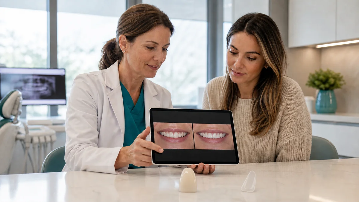
Quick answer: porcelain veneers before and after images can illustrate color, shape, proportion, and visible symmetry, but they cannot prove that veneers are right for your teeth or predict durability. Check whether the photographs are comparable, consented, unfiltered, and taken at a disclosed stage. Then ask about diagnosis, enamel preparation, bite, alternatives, maintenance, and follow-up before deciding.
A dramatic smile comparison can be persuasive in seconds. It can also compress a complex clinical process into two frames. The viewer usually cannot see the examination, radiographs, gum assessment, bite analysis, consent discussion, temporary trial, tooth preparation, laboratory communication, or maintenance plan behind the result. A photograph may document an appearance; it does not independently establish health, suitability, or prognosis.
People searching porcelain veneers before and after often want to know whether their own discoloration, chipped edges, spacing, asymmetry, or worn teeth could change in a similar way. That is a reasonable question, but another person’s images are not a diagnostic test. The same visible concern can arise from different causes, and those causes can call for whitening, bonding, orthodontics, periodontal care, monitoring, a veneer, or a combination rather than one automatic cosmetic treatment.
This guide offers a structured way to read a gallery without mistaking marketing for personalised evidence. It does not diagnose a smile, guarantee an aesthetic result, or assign a fixed lifespan to porcelain. It is designed to help patients ask better questions before healthy tissue is altered and to make the consultation more transparent.
1. porcelain veneers before and after should be read as documentation, not prediction
A fair image pair can show visible changes in tooth length, width, color, surface character, edge position, spaces, and the relationship between teeth and lips. It may help a patient communicate preferences. It cannot tell whether the result was achieved with veneers alone, whether the teeth were healthy beforehand, how much enamel was removed, whether the bite is stable, or how the restorations will behave over time.
The American Dental Association describes a porcelain veneer as a thin custom-made shell placed on the front surface of a tooth. It also distinguishes veneers from crowns, which cover more of the tooth. This distinction matters when interpreting a photograph: a gallery caption should not use “veneers” as a loose label for crowns, composite bonding, whitening, orthodontics, gum treatment, or a mixed rehabilitation.
Think of a gallery as one input among several. It can demonstrate a clinic’s visual style and communication quality. It cannot replace a licensed dentist’s diagnosis, a discussion of relevant options, or valid consent. The useful question is not “Can my teeth look exactly like this?” but “What clinical and photographic information would be needed to decide what is appropriate for me?”
2. An 11-check decision table for evaluating a before-and-after pair
The following table turns visual impressions into practical questions. A single weak point does not prove deception, and a polished image does not prove clinical quality. The aim is to identify missing context before relying on the comparison.
| Check | What to compare | Why it matters | Question to ask |
|---|---|---|---|
| 1. Identity and consent | Clear statement that images are a real, consented case | Protects patient information and discourages borrowed or stock results | Were these photographs used with recorded permission? |
| 2. Same viewpoint | Similar head position, camera angle, crop, and magnification | Angle changes can alter apparent tooth length and symmetry | Are the frames genuinely comparable? |
| 3. Similar lighting | Comparable exposure, color balance, and reflections | Lighting can make shade and surface texture look different | Were filters or selective edits used? |
| 4. Similar lip posture | Relaxed versus stretched smile, lip height, and mouth opening | A changed expression can create a different smile frame | Is the visible improvement partly a pose difference? |
| 5. Treatment label | Veneers alone versus whitening, bonding, orthodontics, or gum care | Combined treatment should not be attributed to porcelain alone | What exactly was performed? |
| 6. Tooth count | Which teeth were treated and which remained natural | Scope affects color matching, preparation, cost, and future care | How many teeth received restorations, and why? |
| 7. Preparation disclosure | No-prep, minimal preparation, or more extensive reduction | The visible front surface does not reveal tissue removal | How much enamel was removed on each tooth? |
| 8. Image timing | Immediate placement versus a later review | Gums, hydration, and adaptation may look different over time | When was the “after” image taken? |
| 9. Bite context | Front view plus relevant side and functional records | A frontal photograph cannot show contact patterns or grinding risk | How was function assessed? |
| 10. Health context | Decay, gum health, cracks, sensitivity, and existing restorations | A beautiful surface cannot prove a healthy foundation | Which conditions were treated before cosmetic care? |
| 11. Follow-up | Immediate appearance versus documented maintenance | Day-of-placement images cannot demonstrate longevity | Is there a review plan and repair pathway? |
Use the table to slow the decision down. An ethical consultation should welcome questions about comparability, treatment scope, alternatives, and limitations. If the only answer is another glamorous image, important clinical information is still missing.
3. Confirm that the images are authentic, consented, and properly described
Dental photographs are patient information. The UK General Dental Council states that using before-and-after images to promote a practice requires the patient’s consent to both taking and promotional use. The patient should understand how the images will be used, consent should be recorded, only necessary information should be released, and permission can be withdrawn. Rules vary by jurisdiction, but the underlying ethical principle is broadly useful: a real patient is not merely marketing material.
A responsible gallery should avoid names, faces, or other identifying details unless the patient has knowingly agreed to that level of disclosure. It should not imply that a stock image, manufacturer image, digitally generated mock-up, or another clinician’s work is an actual treatment result from the publishing clinic. Digital smile simulations should be labelled as simulations rather than “after” photographs.
Ask for clarity on these points:
- Is this an actual treated case from the clinician or team presenting it?
- Did the patient consent to this specific public use?
- Are both frames photographs rather than a photograph paired with a simulation?
- Were brightness, color, tooth contours, or gum lines digitally altered?
- Does the caption name every treatment that materially contributed to the result?
Basic cropping and exposure correction may be part of clinical photography, but adjustments should not manufacture a treatment effect. If editing changes the appearance being judged, the comparison loses evidentiary value.
4. Standardisation matters: angle, light, scale, posture, and timing
Small photographic changes can create a large perceived difference. A camera held slightly higher can change the amount of tooth and gum displayed. A tighter crop can make teeth appear larger. A warmer or cooler white balance can shift perceived shade. A wide smile in one frame and a restrained smile in the other can alter symmetry and tooth display without any dental treatment.
Useful comparisons keep the camera-to-subject relationship, magnification, head orientation, exposure, and lip position as similar as practical. Intraoral views can complement a full-smile image because they show margins, adjacent teeth, and surface details more clearly. They still do not reveal everything beneath a restoration.
Timing should be explicit. An immediate post-bonding photograph records a different stage from a later review after soft tissues have settled and the patient has used the restorations. Teeth can also look optically different when temporarily dehydrated during a long appointment. A gallery should not blur immediate appearance and healed follow-up into the same claim.
5. The foundation comes first: disease and diagnosis are not visible in a gallery
According to the ADA’s patient guidance, dental problems such as decay or gum disease should be treated before veneers are placed, because covering unhealthy teeth may worsen existing problems. The World Health Organization also identifies dental caries and periodontal disease as major oral conditions. These are not side issues in a cosmetic plan; they influence whether a restoration can be cleaned, bonded, maintained, and reviewed safely.
A veneer assessment may include the history of the concern, clinical examination, tooth vitality where indicated, periodontal evaluation, caries risk, existing fillings, cracks, enamel quantity, alignment, bite, and parafunctional habits. Imaging should be selected only when justified by the findings. A social-media image or remote selfie cannot complete those steps.
Before accepting a plan, ask the clinician to name the diagnosis or aesthetic objective for each proposed tooth. “Smile makeover” is not a diagnosis. One tooth may need disease control, another may need no treatment, and another may be suitable for a conservative additive correction. Tooth-by-tooth reasoning reduces the risk of treating the photograph rather than the patient.
6. Ask what happened to the enamel behind the “after” image
Porcelain can look thin and delicate in a photograph, but placement may require removal of tooth structure to create space, define margins, correct position, or avoid an over-contoured result. The ADA notes that veneer treatment is not reversible when enamel is removed. The NHS similarly explains that the front of the tooth is drilled away a little for some veneers, while not all veneer approaches require the same preparation.
“No-prep” is not a universal promise of no biological cost. A genuinely additive approach may be suitable in selected anatomy, but placing extra thickness on an already prominent or crowded tooth may compromise contour, emergence, or cleaning. Conversely, a tooth that is discolored, rotated, fractured, or already restored may need a different design. The required preparation is a clinical decision, not a package label.
A useful plan should explain:
- which surfaces and teeth require preparation;
- whether margins are expected to remain primarily in enamel;
- why the proposed reduction is necessary for contour, strength, or color control;
- what sensitivity, repair, and replacement implications were discussed;
- whether a less invasive path can meet the same goal.
7. Compare conservative alternatives before selecting porcelain
The 2024 AWMF S3 guideline on direct composite restorations recommends minimally invasive direct procedures that preserve tooth structure for anterior shape corrections whenever possible, with indirect ceramic veneers as an alternative. FDI’s minimal intervention policy likewise supports preserving healthy tooth structure and avoiding unnecessary removal. These principles do not make composite automatically better; they require the least destructive option capable of meeting the agreed objective.
Alternatives depend on the underlying concern. Whitening may address generalized color without changing shape. Orthodontics may move teeth rather than mask position. Direct composite bonding may add or repair form with a different maintenance profile. Contouring may help a limited edge discrepancy in selected cases. Periodontal treatment may be necessary before any cosmetic restoration. Sometimes monitoring or no treatment is the safest answer.
Comparison should cover biological cost, expected maintenance, reparability, color behavior, appointment sequence, and the effect on future options. Porcelain may be appropriate when the indication, tooth substrate, design, and patient priorities align. Its presence in an attractive gallery is not itself an indication.
8. Use mock-ups and provisional stages to test a plan before final porcelain
Patients often react to a final image as a whole but make decisions tooth by tooth. A diagnostic design, physical mock-up, provisional restoration, or other reversible preview may help assess length, width, speech, lip support, tooth display, and overall style before final manufacture. Digital simulation can support communication, but it should be identified as a planning illustration rather than a guaranteed result.
A preview is most useful when it invites specific feedback. “Do you like it?” is less informative than asking whether the edge length feels comfortable, whether the central incisors dominate too much, whether the smile looks too uniform, and whether speech or lip closure changes. The clinician can then discuss which requests are compatible with anatomy and function.
Even a well-made trial has limits. Material, translucency, surface texture, and gum response may differ from the definitive restoration. Its purpose is to reduce uncertainty and improve communication, not to promise an exact photographic duplicate.
9. Judge naturalness through more than whiteness
Very white teeth can dominate a phone screen, but natural-looking results involve more than shade. Tooth proportions, incisal edge position, surface texture, translucency, line angles, contact areas, gingival levels, and the transition to untreated teeth all affect perception. A result may be technically bright yet visually flat, bulky, or disconnected from the patient’s face.
Ask whether the plan aims for a uniform cosmetic style or a more individual appearance. Neither preference is inherently correct, but it should be explicit. The clinician and laboratory need to understand how much variation, texture, translucency, and asymmetry the patient finds acceptable.
Color matching also depends on scope. Treating one or two teeth next to natural enamel can require a different strategy from restoring a broader visible segment. Whitening natural teeth before shade selection may be considered in some plans, while existing crowns or fillings may not respond like enamel. A caption should disclose relevant combined care rather than credit every change to veneers.
10. A frontal smile cannot show bite, grinding, or functional risk
The ADA advises that veneers may not be a good choice for some people who clench or grind their teeth or have a deep overbite. A frontal photo cannot reveal all functional contacts, guidance patterns, muscle symptoms, wear behavior, or the direction of forces on the veneer edges. Those findings require clinical assessment.
Function matters because the restoration becomes part of a moving system. Tooth position, opposing teeth, missing teeth, edge-to-edge contacts, and habits such as nail biting or chewing hard objects can influence design and maintenance. A protective appliance may be discussed for selected patients, but it does not make an unsuitable plan suitable or eliminate every risk.
Tell the dentist about jaw discomfort, morning muscle fatigue, previous chips, frequent debonding, sports, instrument habits, and nighttime grinding. These details may change material, preparation, tooth count, sequence, or whether veneers are advisable at all.
11. Separate immediate appearance from health, comfort, and longevity
An immediate “after” photograph may show clean margins and an attractive smile, yet it cannot establish long-term comfort, pulp health, gum stability, bonding durability, color integration, or patient satisfaction. The ADA notes that veneers can chip, crack, wear, loosen, or require repair or replacement. That is not a prediction that a specific restoration will fail; it is a reason to plan maintenance and future care before starting.
Ask whether the gallery includes review photographs and how those images are labelled. A later image can add context, but it still represents one patient with a particular starting condition, design, behavior, and maintenance history. It should not be converted into a universal lifespan claim.
Meaningful outcomes include more than appearance:
- comfort at rest and during chewing;
- cleanable margins and stable gum tissues;
- acceptable speech and tooth display;
- manageable sensitivity, if any;
- a documented pathway for review, repair, or replacement.
12. Understand scope: veneers alone may not explain the visible change
Some transformations involve staged care. Orthodontics may first improve tooth position and reduce the amount of restorative masking needed. Whitening may establish a lighter background shade. Gum therapy may improve health before definitive margins are planned. Composite may be used on selected teeth while porcelain is used on others. Old restorations may need separate management.
Ask for a written sequence showing what happens first, which steps are optional, and what conditions could change the plan. This is especially important when several clinicians or a dental laboratory are involved. The final caption may fit in one line; the actual care pathway should not.
The number of veneers should follow visible need, tooth condition, smile dynamics, and color-matching strategy, not a standard bundle. More treated teeth can create broader control over appearance, but they also expose more teeth to intervention and future maintenance. A tooth that does not need treatment should not be included merely to reach a package count.
13. Maintenance belongs in every porcelain veneers before and after discussion
Veneers do not prevent future decay or gum disease. The ADA recommends brushing with fluoride toothpaste, cleaning between teeth, avoiding harmful object-biting habits, and using appropriate protection for activities that could injure the mouth. Recall intervals and professional maintenance should be tailored to oral health, restoration design, disease risk, and clinician findings.
Ask how the margins should be cleaned, whether particular flossing or interdental techniques are needed, and whom to contact if a veneer feels mobile, rough, high, or uncomfortable. Clarify how emergency review, minor repair, rebonding, remaking, and management of the underlying tooth are handled. Maintenance responsibilities should be written clearly when treatment is provided away from home.
No restoration should be described as permanent in the sense of never needing care. Future work may be necessary because of wear, fracture, debonding, gum change, disease, trauma, or changing aesthetic preferences. Consent is stronger when that future pathway is discussed before preparation.
14. Planning veneers in Turkey: continuity matters as much as the gallery
International care adds logistics to an already detailed clinical decision. Travel dates should not force diagnosis, disease control, laboratory steps, bonding, bite adjustment, or review into an unsafe timetable. A patient may need additional assessment or a revised plan after examination; a clinic should be willing to say so.
When discussing veneer care with Redent Klinik, provide a dental history, current symptoms, medications, allergies, relevant previous treatment, and any available records. Remote photographs can support initial communication, but the final plan should follow an in-person examination and justified diagnostics. The clinic should explain which parts of a preliminary proposal may change.
Before booking travel, request a written outline covering appointment stages, provisional care where relevant, what happens if more treatment is found, and how post-treatment questions will be managed. The Redent Klinik contact team can help organise communication, but clinical suitability and timing remain decisions for the treating dental team.
15. Gallery and sales red flags that justify a pause
Attractive photographs and ethical care can coexist. Concern arises when images are used to suppress rather than support questions. Pause when a provider treats visual similarity as a guarantee, offers a definitive plan from selfies alone, refuses to discuss alternatives, or presents preparation as universally reversible.
Other warning signs include:
- before and after frames with radically different lighting, angles, filters, or expressions;
- no disclosure that whitening, orthodontics, gum care, bonding, or crowns contributed;
- identical-looking images appearing under unrelated clinics or accounts;
- pressure to choose a fixed tooth count before examination;
- claims of painless, risk-free, maintenance-free, or guaranteed treatment;
- care offered by an unlicensed “veneer technician” without dental diagnosis;
- no written plan for consent, records, review, or complications.
The ADA specifically cautions against veneer services that remove the licensed dentist from diagnosis and treatment. Cosmetic dentistry remains dentistry. A healthy-looking marketing image does not make unsupervised treatment safe.
16. A consultation checklist for turning images into informed consent
The GDC’s consent standard requires relevant options and possible costs to be explained before treatment, with consent treated as ongoing communication rather than a signature alone. Even outside the UK, this is a useful model for a cosmetic consultation. The patient should understand what is proposed, why, what alternatives exist, what may change, and what future care could involve.
Bring a small selection of reference images and describe the features you like rather than requesting a duplicate smile. Then ask:
- What is the diagnosis or objective for each tooth?
- Which teeth can remain untreated?
- How much enamel is expected to be removed, and why?
- Could whitening, orthodontics, composite, contouring, or no treatment meet the goal?
- How will bite, grinding, gum health, and decay risk be assessed?
- Can a mock-up or provisional stage test shape and function?
- What is included in the treatment scope and what is separate?
- What maintenance, review, repair, and replacement pathways are available?
- How are clinical photographs stored, and is public use optional?
Consent to treatment and consent to marketing photography are separate decisions. A patient can accept care without agreeing to public image use. Refusing promotional use should not reduce the quality of treatment or follow-up offered.
17. When symptoms need assessment instead of cosmetic comparison
Do not use a gallery to self-select cosmetic treatment when there is pain, swelling, spontaneous bleeding, a loose tooth, a recent injury, a broken restoration, or a veneer that feels mobile. These findings may need prompt professional assessment. Severe swelling, difficulty breathing or swallowing, uncontrolled bleeding, or significant facial trauma can require urgent care according to local emergency pathways.
After veneer placement, contact the treating dentist if the bite feels persistently high, a margin catches, sensitivity is severe or worsening, a restoration moves, or the gum remains painful. A photograph sent to the clinic may help triage, but it does not replace examination when symptoms or function are involved.
Frequently asked questions about porcelain veneers before and after
Can before-and-after photos prove that a dentist is good at veneers?
They can show selected visible work and may reveal a clinic’s aesthetic style, but they cannot independently prove diagnosis quality, preparation depth, bonding, function, health, or durability. Look for standardised images, full treatment disclosure, informed consent, and a clinician who explains limitations and alternatives.
How can I tell whether veneer photos are edited?
Compare lighting, background color, skin tone, reflections, tooth edges, gum contours, crop, and sharpness. Ask directly whether filters, retouching, or digital simulation were used. Ordinary exposure adjustment is different from changing the feature being evaluated; material edits should be disclosed.
Should the “before” and “after” photos use the same lighting?
Lighting and white balance should be as comparable as practical because both affect perceived shade and texture. Angle, scale, head position, and lip posture also matter. Exact duplication may be difficult, but large unexplained differences reduce the comparison’s value.
Does an immediate after photo show the final result?
It shows an early stage. Gum tissues, hydration, comfort, bite adaptation, and the patient’s perception may evolve. Ask when the image was taken and whether a later clinical review occurred. Neither immediate nor later photographs can guarantee future performance.
Can porcelain veneers fix crooked teeth?
Veneers can mask selected visible irregularities, but they do not move roots or correct every bite problem. Masking position may require preparation or create contour challenges. Orthodontics, bonding, combined care, or no treatment may be more conservative depending on the diagnosis.
Are no-prep veneers always reversible?
No. The label does not prove that no enamel adjustment, etching, margin finishing, or future intervention will occur. Suitability depends on tooth position, volume, color, and desired contour. Ask for a tooth-by-tooth preparation plan rather than relying on a package name.
Will my result look exactly like a reference photo?
No clinician should guarantee an exact copy. Tooth substrate, lip dynamics, gums, face, bite, material thickness, and personal perception differ. Reference images are best used to discuss preferences such as brightness, texture, edge shape, and degree of uniformity.
How many teeth need porcelain veneers?
There is no universal number. The visible smile, treatment objective, health of each tooth, color matching, existing restorations, and alternatives should guide scope. Treating more teeth simply to complete a package can expose sound teeth to unnecessary intervention.
Can I consent to treatment but refuse public photography?
Yes. Clinical records may include necessary photographs under applicable care and privacy rules, but promotional use requires a separate, properly explained basis. Ask how images will be stored and used. A refusal of marketing use should not affect treatment quality.
What should I do if a veneer chips or feels loose?
Avoid biting with the affected area, keep any detached fragment or restoration safe, and contact a dentist. Do not glue it yourself. Urgency depends on pain, sharp edges, exposed tooth, trauma, and aspiration risk, so obtain professional triage.
Final perspective: use the image to start questions, not end them
The most reliable way to use porcelain veneers before and after is to treat each pair as a prompt for verification. Confirm that the images are authentic and comparable. Ask what treatment was actually performed, how much tissue was altered, when the result was photographed, what alternatives were considered, and how health and function were assessed.
A visually appealing result can be meaningful without becoming a promise. Your safest plan is the one grounded in an examination, a clear diagnosis or objective, conservative option comparison, valid consent, realistic expectations, and a workable maintenance pathway. The goal is not to reproduce another person’s photograph. It is to make an informed decision that respects your own teeth, priorities, and future care.
Sources and evidence
- American Dental Association: Official website
- American Dental Association, MouthHealthy: Veneers
- NHS: Dental treatments, including veneers
- General Dental Council: Obtain valid consent
- General Dental Council: Patient information and before-and-after image consent
- AWMF S3 Guideline: Direct composite restorations in permanent anterior and posterior teeth
- FDI World Dental Federation: Minimal Intervention Dentistry
- World Health Organization: Oral health fact sheet