
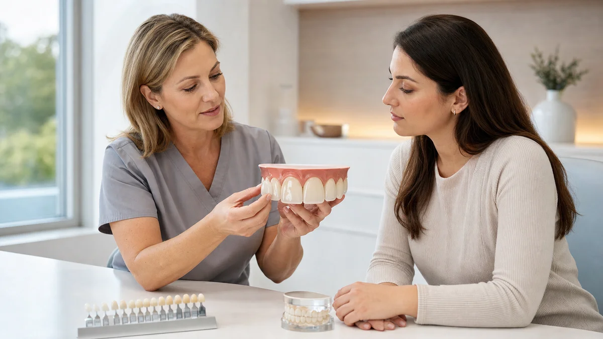
Quick answer: Important porcelain veneers risks include irreversible enamel reduction, temporary or persistent sensitivity, fracture, chipping, debonding, decay at exposed margins, gum irritation, bite problems and an eventual replacement cycle. Risk is not identical for every patient. It rises when active disease, limited enamel, grinding, unrealistic shape changes or poor bonding conditions are ignored. Careful diagnosis, conservative planning and maintenance reduce risk but cannot remove it.
Porcelain veneers can improve the color, contour, symmetry and proportions of selected front teeth. They are thin ceramic restorations bonded mainly to the visible surfaces of prepared teeth. When the indication, preparation, ceramic, bonding and bite are well matched, clinical research generally reports favorable outcomes. That evidence does not mean that veneers are reversible, permanent or free from complications.
Risk discussions are often distorted in two directions. Marketing may present veneers as a harmless cosmetic layer that lasts forever. Alarmist posts may imply that every veneer destroys a tooth or inevitably causes root canal treatment. Neither framing helps informed consent. The relevant questions are which teeth actually need treatment, how much sound enamel can be preserved, what biological and technical events are plausible, and what happens when a veneer eventually needs repair or replacement.
This guide organizes risk by the stage at which it arises: diagnosis, preparation, bonding, early adaptation and long-term maintenance. It uses public guidance from the American Dental Association and the US National Institute of Dental and Craniofacial Research alongside peer-reviewed clinical reviews. It is educational and cannot determine whether a particular tooth is suitable for veneers.
porcelain veneers risks start before a tooth is prepared
The highest-impact error is often a wrong indication, not a ceramic defect. A veneer changes the surface and shape of a tooth; it does not treat active decay, gum disease, an infected pulp, an unstable bite or a root fracture. It may camouflage a visual concern while the underlying cause continues. Every cosmetic plan should therefore begin with a dental and periodontal examination, appropriate imaging when clinically indicated, and a review of function and health history.
The dentist should identify the patient’s primary concern in plain language. Whitening may address generalized color. Composite bonding may repair a small chip. Orthodontic treatment may correct position rather than cover it. A direct restoration may treat localized decay. Monitoring may be reasonable when a harmless variation does not require intervention. A veneer is justified only when its expected benefit exceeds the biological cost and less invasive choices do not meet the agreed objective.
A mock-up or trial smile can help test length, width, speech and lip support before enamel is removed. Digital simulations can support communication, but they are not guarantees. Screen images do not reproduce ceramic translucency, gum response, bite forces or healing. Consent should be based on clinical limitations as well as the proposed appearance.
Decision table: 12 risks, warning signs and controls
| Risk | What can increase it | What a patient might notice | Risk-control discussion |
|---|---|---|---|
| 1. Unnecessary irreversible treatment | Cosmetic pressure, no diagnosis, no alternatives review | Healthy teeth are proposed for broad treatment without a problem list | Confirm the smallest treatment that solves the stated concern |
| 2. Excessive enamel or dentin exposure | Major color or position change, over-contour correction, poor planning | More preparation than expected or a change from veneer to crown | Use a diagnostic mock-up and tooth-specific preparation plan |
| 3. Sensitivity or pulpal symptoms | Deep preparation, cracks, existing restorations, bonding trauma | Cold sensitivity, spontaneous pain or pain on biting | Assess pulp health, preserve enamel and review persistent symptoms |
| 4. Debonding | Limited enamel, contamination, unfavorable forces or preparation | Veneer feels mobile or detaches | Evaluate bonding substrate, isolation, occlusion and cement protocol |
| 5. Fracture or chipping | Grinding, edge-to-edge bite, trauma, thin or unsupported ceramic | Rough edge, visible line, missing fragment | Plan thickness and bite; discuss protection and repair limits |
| 6. Decay at or near a margin | High sugar frequency, dry mouth, plaque, exposed root or poor fit | Darkness, sensitivity, odor, food trapping or no early symptom | Control decay risk and keep margins accessible for hygiene |
| 7. Gum inflammation or recession | Over-contour, deep margins, plaque, active gum disease | Bleeding, swelling, tenderness, uneven gum line | Stabilize gums first and design cleanable emergence contours |
| 8. Bite discomfort or wear | Incorrect contacts, parafunction, unpolished adjustments | Tooth hits first, jaw fatigue, opposing tooth feels rough | Record function, verify movements and polish adjusted ceramic |
| 9. Color or shape dissatisfaction | Unrealistic preview, inadequate shade communication, opacity limits | Too white, opaque, bulky, long or asymmetric appearance | Approve provisionals or try-in and define acceptable limitations |
| 10. Repair or matching difficulty | Single later failure, discontinued material, changing natural teeth | Replacement looks different from neighboring veneers | Keep records and discuss repair versus remake before treatment |
| 11. Repeated replacement cycle | Young age, extensive initial preparation, recurrent complications | Each remake may need new preparation or broader treatment | Consider lifetime maintenance, not only the first placement |
| 12. Unsafe unlicensed care | Direct-to-consumer kits or a non-dentist placing restorations | No examination, sterilization assurance or complication pathway | Use a licensed dentist accountable for diagnosis and follow-up |
These porcelain veneers risks are not a prediction that all events will happen. The table is a consent framework. One patient may have excellent enamel and a low-risk bite but significant color limitations; another may have healthy-looking front teeth with active grinding and little enamel available for bonding. The same veneer design cannot carry the same risk in both mouths.
Risk 1: enamel reduction is generally irreversible
Traditional porcelain veneer preparation removes a controlled amount of surface tooth structure to create space and a defined finish line. Enamel does not grow back. Even a conservative preparation can mean that the tooth will continue to need a bonded restoration or another covering restoration in the future. This is why elective treatment of healthy teeth deserves a higher consent standard than a reversible cosmetic product.
Preparation depth varies by tooth position, existing shape, discoloration, ceramic choice and desired change. A tooth that protrudes may require more reduction to avoid a bulky result. Masking a very dark tooth may require ceramic thickness or opacity that conflicts with a minimal preparation. Closing a large space can create over-wide teeth unless orthodontics or a combined plan is considered.
Research has repeatedly examined the bonding substrate. Enamel generally provides a favorable and durable surface for adhesive ceramic veneers. Reviews and clinical studies have associated greater dentin exposure with less favorable outcomes than preparations largely confined to enamel. That does not mean every small area of dentin leads to failure; it supports planning that preserves enamel whenever possible and honest discussion when it cannot be preserved.
“No-prep” does not automatically mean no risk or complete reversibility. Adding ceramic without creating space can produce over-contoured teeth, ledges, altered speech or gum irritation in an unsuitable case. Some no-prep designs still involve cleaning, etching or minor modification of enamel. The dentist should state exactly what will be changed rather than relying on a marketing label.
Risk 2: sensitivity, pain and possible pulp treatment
Temporary sensitivity to cold or air can occur after tooth preparation and bonding. The tooth has been manipulated, and the bite and contacts have changed. Mild symptoms may settle, but spontaneous pain, worsening night pain, prolonged thermal pain, swelling or significant pain on biting requires assessment. Patients should not be told that every painful response is normal or that all sensitivity means a root canal is inevitable.
Pulp risk can be influenced by preparation depth, previous decay, large restorations, cracks, trauma and the original health of the tooth. Heat, dehydration or contamination during treatment can also matter. A veneer is thin, but the procedure still involves a living tooth. A pre-treatment sensibility examination and radiograph may be appropriate when history or findings raise concern.
If the pulp later becomes irreversibly inflamed or infected, root canal treatment may be necessary. Access can sometimes be made through the back of the tooth without removing the veneer, but the restoration may be damaged or need replacement. No clinician can guarantee that a prepared tooth will never require endodontic care.
Risk 3: debonding can expose a prepared surface
Porcelain veneers rely on an adhesive interface among ceramic, resin cement and tooth. Debonding may occur when this system is compromised by insufficient enamel, unfavorable preparation geometry, contamination, an incompatible surface-treatment protocol or excessive forces. A detached veneer should be kept safely and assessed; a patient should not use household glue or attempt to rebond it.
Rebonding may be possible when the veneer is intact, the tooth is healthy and surfaces can be properly cleaned and retreated. It is not automatically the best choice. The clinician must check why it detached, whether the fit remains accurate, whether decay or fracture is present and whether occlusal forces need correction. Repeating the same bonding steps without addressing the cause may repeat the event.
Bonding is technique-sensitive. Isolation from saliva and blood, correct ceramic conditioning, enamel and dentin management, compatible materials, complete seating and careful removal of excess cement all influence the interface. Patients can ask which material is being used, but a brand name alone does not verify correct execution.
Risk 4: porcelain can fracture, chip or develop cracks
Dental ceramics are strong under intended conditions but are also brittle. A veneer can chip at an edge, crack or fracture after impact, repeated loading or biting a hard object. Thin unsupported areas, inadequate ceramic thickness, internal defects, poor fit and unfavorable bite contacts may contribute. A small rough area may be polishable or repairable with composite; a major fracture often requires replacement.
Systematic reviews describe fracture and debonding among the important technical complications of porcelain laminate veneers. Reported rates vary because studies include different materials, preparation designs, patient groups, follow-up periods and definitions of success. A pooled survival number cannot predict one person’s result or justify a lifetime guarantee.
After any facial or dental trauma, a fractured veneer may accompany a fractured tooth or root. The visible ceramic is only one part of the diagnosis. Pain, mobility, bleeding, a changed bite or a darkening tooth should be evaluated promptly.
Risk 5: decay can still develop around a veneer
A veneer covers only part of a tooth. The back surface, contact areas, root surfaces and margins remain exposed to the oral environment. Plaque bacteria can contribute to decay when frequent sugars, dry mouth, inadequate cleaning and susceptible tooth surfaces combine. A well-fitting veneer does not make the tooth cavity-proof.
Early decay may cause no symptoms. NIDCR notes that tooth decay can progress before pain is present. This is why routine dental review remains important after cosmetic treatment. Clinically indicated radiographs, visual examination and risk assessment can identify problems that a mirror or photograph does not show.
Decay at a margin may sometimes be managed with a localized restoration, but access, extent and appearance influence the choice. More extensive decay can require veneer removal, a new veneer, a crown or endodontic treatment. If a tooth is no longer restorable, extraction may eventually be discussed. Good hygiene reduces risk but cannot guarantee that recurrent decay will never occur.
- Brush twice daily with fluoride toothpaste using a gentle gumline technique.
- Clean between veneered teeth with a method suited to the contact and gum space.
- Limit frequent sugary drinks and snacks rather than relying only on surface cleaning.
- Tell the dental team about dry mouth, new medicines, reflux or eating disorders.
- Attend risk-based examinations and professional cleaning rather than waiting for pain.
Risk 6: gum inflammation, recession and margin exposure
Healthy gums are part of the visual result. Active gingivitis or periodontitis should be controlled before definitive veneers because swelling and bleeding can distort margin position, make isolation difficult and produce an unstable reference for design. NIDCR describes plaque as a central cause of gum inflammation and notes that tartar requires professional removal.
Veneer contours that are bulky or overhanging can retain plaque and make cleaning difficult. Margins placed unnecessarily deep below the gum may complicate hygiene and tissue response. Excess bonding cement left near the margin can also irritate tissues. The goal is a smooth, cleanable transition consistent with biological space and appearance.
Gum levels can change over time because of periodontal disease, brushing trauma, natural tissue changes, tooth movement or orthodontic treatment. Recession may expose a darker root or the veneer-tooth junction, creating a visible line even when the ceramic remains intact. Replacing a veneer does not by itself treat the cause of recession.
Persistent bleeding, swelling, bad taste, tenderness or increasing recession warrants an examination. Aggressive brushing or unverified bleaching around the margin can worsen irritation. Treatment may involve hygiene coaching, periodontal care, contour correction, cement removal, repair or replacement depending on the diagnosis.
Risk 7: bite problems and bruxism change technical risk
Veneers participate in biting and jaw movements, especially when they extend over incisal edges. The dentist should evaluate how front and back teeth contact in closure and movement. If one veneer contacts too early, the patient may notice a tooth that “hits first,” tenderness, jaw fatigue or a sense that the bite is uneven.
Bruxism includes clenching or grinding behaviors that may occur during sleep or while awake. Clinical research has associated bruxism activity with a higher risk of ceramic failure in veneer patients. A protective splint may be discussed for selected patients, but it cannot guarantee prevention and must itself be monitored for fit and wear.
Signs such as tooth wear, fractures, jaw-muscle tenderness, tongue or cheek changes and a history of broken restorations inform planning. The team may modify design, choose another material, stage treatment or recommend managing function before elective veneers. A patient should not conceal grinding because of concern that treatment will be refused; it is central safety information.
Adjusted porcelain surfaces should be finished and polished appropriately. A rough ceramic edge can feel irritating and may interact unfavorably with opposing teeth. Simply grinding a high spot without restoring a smooth surface is incomplete care.
Risk 8: color, opacity, proportion and symmetry may disappoint
Porcelain does not whiten after placement. Natural neighboring teeth may change color with age, staining or bleaching while veneers remain relatively stable. A later replacement made from a different ceramic batch or by another laboratory may not match perfectly. Shade records, photographs and laboratory communication can help but do not eliminate visual variation.
Masking a very dark tooth can require opacity, which may reduce the depth and translucency patients expect from natural enamel. A very white shade can make adjacent natural teeth look darker. Multiple identical veneers can appear artificial if natural texture, incisal variation and tooth-specific proportions are ignored.
Tooth width, length and position affect speech and lip movement. Closing large gaps solely by adding width can create broad teeth. Lengthening edges can alter sounds and bite contacts. Provisional restorations or a mock-up allow the patient and clinician to test these features before final ceramic is bonded, although provisional material will not duplicate every optical detail.
Consent should define the acceptable range, not promise perfection. Facial asymmetry, gum levels and natural tooth position may limit symmetry without orthodontic or periodontal treatment. A responsible clinician can explain when an additional procedure is optional, necessary or not justified.
Risk 9: replacement and repair can become more complex over time
A veneer may eventually need polishing, repair, rebonding or replacement. Removal must be performed carefully because the boundary between ceramic, cement and tooth can be difficult to judge. Additional tooth structure may be lost during removal or when new decay and fractures are treated. A later restoration can therefore require a broader preparation than the first.
Small chips may be smoothed or repaired with resin composite, but color and surface texture may not match perfectly. Debonded intact veneers can sometimes be rebonded after diagnosis. A fractured or poorly fitting veneer usually requires a remake. If insufficient tooth structure remains, a crown may be discussed, although a crown is not an automatic consequence of every replacement.
Age matters because a younger patient has more years in which replacements may be needed. Long-term porcelain veneers risks therefore include the cumulative burden of monitoring and remakes, not only an early chip or debond. The initial quote is only one part of lifetime burden. Consent should include expected review, maintenance, repair access and the financial possibility of future replacement without presenting a fixed lifespan.
Risk 10: material labels can hide meaningful differences
“Porcelain veneer” is a broad phrase. Feldspathic porcelain, leucite-reinforced ceramic, lithium disilicate and other ceramic systems have different optical and mechanical properties. The ADA explains that indirect restorative materials differ in composition, fabrication, properties and indications. Material selection should follow the clinical design rather than a universal ranking.
Very thin feldspathic porcelain may provide optical layering in a suitable enamel-bonded case. Pressed or milled glass-ceramics can offer different strength and fabrication options. Zirconia veneers exist, but evidence and bonding behavior should not be assumed identical to silica-based ceramics. A product advertised as “the strongest” may not be the most translucent, bondable, repairable or conservative choice for a specific tooth.
Ask whether the restoration is monolithic or layered, how the internal surface will be treated, which cement system is planned and how thickness relates to the desired shade. Patients do not need to direct laboratory chemistry, but the clinician should be able to explain why a material fits the indication and what evidence limitations remain.
Risk 11: unlicensed and direct-to-consumer veneer care
The ADA warns that veneer services from unlicensed individuals can expose patients to infection, nerve damage and placement over unhealthy teeth. A dental veneer is not merely a decorative accessory. Diagnosis, preparation, bonding, bite adjustment and management of complications are dental acts that require accountability, infection control and professional records.
Mail-order or direct-to-consumer products may use self-taken impressions or removable cosmetic shells. These are not equivalent to clinician-planned bonded porcelain veneers. A device that covers teeth can trap plaque, alter the bite, irritate gums or conceal decay. Online images cannot show pulp health, cracks, periodontal support or occlusal function.
Warning signs include no request for a dental examination, no licensed dentist identified, no medical history, no sterilization explanation, pressure to pay before diagnosis and no urgent-care pathway. Do not allow permanent cement, etching chemicals or rotary instruments to be used by an unlicensed provider.
Risk 12: active disease and health factors can undermine the plan
Decay and gum disease must be assessed and controlled before elective veneers. Dry mouth can increase decay risk and may be associated with medicines, medical conditions or radiation therapy. Smoking affects gum health and healing. Reflux and frequent acidic exposure can erode tooth surfaces. Eating disorders can involve acid exposure, nutrition and psychological needs that require sensitive multidisciplinary care.
Pregnancy does not make all dental care unsafe, but elective cosmetic timing, radiographs, medicines and gum inflammation should be discussed with the dentist and relevant clinician. Anticoagulants, immune-modifying medicines and other prescriptions may affect invasive supporting procedures. Never stop prescribed medicine based on a cosmetic treatment article.
Expectations and decision pressure are also safety factors. A patient seeking an exact celebrity smile, complete facial transformation or treatment during a personal crisis may benefit from more time, a mock-up or a second opinion. Consent is not meaningful when the patient believes normal variation is a defect or that veneers will solve unrelated confidence and life concerns.
How clinicians can reduce porcelain veneers risks
Risk reduction begins with a tooth-by-tooth plan and ends with long-term review. It is not a promise that a complication cannot occur. A robust pathway should include:
- Diagnosis: chart decay, restorations, cracks, pulp findings, gum health and bite before cosmetic design.
- Alternative review: compare whitening, bonding, orthodontics, contouring, repair and monitoring where relevant.
- Preview: use photographs, wax-up, mock-up or provisionals to evaluate proportion, speech and lip support.
- Conservative preparation: preserve sound enamel and avoid converting a veneer indication into unnecessary full coverage.
- Material-design match: select ceramic, thickness, translucency and edge design for the individual tooth and bite.
- Controlled bonding: use appropriate isolation, surface treatment, cementation and complete excess-cement removal.
- Functional verification: check static and moving contacts and polish adjusted ceramic.
- Maintenance plan: define hygiene tools, review timing, grinding management and the response to a chip or debond.
Before preparation, ask the dentist to distinguish what is necessary for health, what is necessary to achieve the agreed cosmetic design, and what is optional. That separation makes consent clearer and helps prevent treatment from expanding unnoticed.
Alternatives when veneer risk outweighs benefit
No treatment or monitoring is a legitimate option when teeth are healthy and the concern is minor. Professional whitening can address suitable color concerns without changing tooth shape, though it has its own indications and sensitivity considerations. Composite bonding can change localized shape and may be more repairable, but it can stain, chip or wear and still requires sound technique.
Orthodontic treatment can reposition teeth and improve spacing or bite rather than disguising position with ceramic. It requires time and retention and is not risk-free, but may preserve more tooth structure. Small enamel contour changes can sometimes improve symmetry within safe limits. Existing restorations may be repaired or replaced selectively rather than covering every visible tooth.
A crown is more extensive than a veneer and is generally considered when the tooth needs broader structural coverage, not simply because a veneer is technically challenging. Extraction and implants are not cosmetic substitutes for restorable healthy teeth. The safest alternative solves the actual problem with the least justified biological cost.
Planning porcelain veneers with Redent Klinik in Turkey
Discussing porcelain veneers risks before dental travel requires enough time for diagnosis, planning, any disease control, preparation, laboratory work, trial, bonding and review. Remote photographs can help organize a preliminary discussion but cannot determine enamel thickness, pulp status, cracks, periodontal health or bite. The final plan may change after an in-person examination and clinically indicated imaging.
Patients can review the Redent Klinik English website and use the Redent Klinik contact page to share questions and relevant records. Ask who performs each stage, which ceramic and laboratory are proposed, how many teeth are truly indicated, and how complications will be managed after returning home.
Allow time to evaluate a mock-up or provisionals rather than approving irreversible changes under travel pressure. Obtain copies of photographs, radiographs, tooth chart, material information and treatment notes. Arrange a local dentist for routine maintenance and urgent review. No clinic should guarantee a lifetime result or diagnose veneer suitability from social-media images alone.
When symptoms need prompt assessment
Contact a dentist promptly if a veneer becomes mobile, fractures, leaves a sharp edge, or is associated with worsening sensitivity, spontaneous pain, pain on biting, swelling, pus or a changed bite. Keep a detached fragment or veneer in a clean protective container. Do not glue it or continue biting on an unstable restoration.
Facial swelling, fever with spreading dental infection, difficulty swallowing or difficulty breathing may require urgent or emergency care. Persistent gum bleeding, a non-healing ulcer, marked recession or an unexplained color change in the underlying tooth also deserves examination. Painkillers and cosmetic repair kits do not diagnose the cause.
Frequently asked questions about porcelain veneers risks
What are the most important porcelain veneers risks?
The main categories are irreversible tooth reduction, sensitivity or pulpal problems, debonding, fracture, decay, gum inflammation or recession, bite issues, aesthetic dissatisfaction and future replacement. Individual risk depends on diagnosis, enamel, bite, hygiene and technique.
Do porcelain veneers ruin healthy teeth?
Properly indicated conservative veneers do not inevitably ruin teeth, but preparation is usually irreversible and changes future restorative needs. Covering healthy teeth without a justified goal or removing excessive structure creates avoidable harm. Alternatives and preparation extent should be explicit before consent.
Can porcelain veneers cause permanent sensitivity?
Temporary sensitivity can occur. Persistent or worsening symptoms are less common but may reflect bonding issues, bite trauma, cracks or pulp inflammation. The tooth should be examined rather than assuming the symptom will resolve or that root canal treatment is automatically required.
Can teeth decay under veneers?
Yes. The tooth remains biological and much of it is uncovered. Decay can develop at margins, between teeth, on roots or on the back surface. Fluoride toothpaste, interdental cleaning, diet control and risk-based dental review remain necessary.
Can gums recede after veneers?
Gum levels can change from plaque inflammation, periodontal disease, brushing trauma, tooth position, tissue type or aging. Recession may expose a margin or root. Cleanable contours, healthy gums before treatment and maintenance can reduce risk but cannot guarantee stable tissue forever.
Do porcelain veneers fall off?
Debonding is a recognized complication but is not expected as a routine event. Enamel availability, isolation, surface treatment, preparation, fit and bite affect retention. A detached veneer requires professional diagnosis before rebonding or replacement.
Are no-prep veneers risk-free and reversible?
No. They may be suitable for selected small or recessed teeth, but adding ceramic without space can create bulk, poor contours or bite problems. Enamel may still be conditioned or minimally modified. Removal without any tooth effect cannot be guaranteed.
Is porcelain safe for people who grind their teeth?
Grinding can increase technical risk. It does not automatically exclude every patient, but the pattern and severity should be assessed. Design changes, management of contributing factors and a protective appliance may be discussed without promising complete protection.
Can a chipped veneer be repaired?
Small chips may sometimes be polished or repaired with composite. Larger fractures, poor fit, color concerns or underlying tooth damage may require replacement. A repair may remain visible and its longevity cannot be guaranteed.
Will veneers stay the same color as natural teeth?
Ceramic is relatively color stable, but neighboring natural teeth can stain or be whitened while the veneer does not respond the same way. Gum recession and cement or tooth color can also alter appearance. Shade planning should consider untreated teeth and future whitening.
How many teeth should receive veneers?
There is no safe standard number. The smile display, tooth condition, symmetry, color transition, bite and patient goal matter. Treating more teeth only for uniformity increases irreversible treatment and future maintenance, so each tooth needs a separate indication.
Should I get veneers from a veneer technician?
No unlicensed provider should diagnose, prepare or permanently bond dental restorations. The ADA warns about infection and nerve-damage risks from unlicensed veneer services. Choose a licensed dentist responsible for examination, treatment, infection control and follow-up.
Do porcelain veneers last forever?
No. Clinical studies show favorable survival in selected cases, but fractures, debonding, decay, endodontic treatment and aesthetic changes occur. Research designs and follow-up vary. Patients should plan for maintenance and possible repair or replacement rather than rely on a fixed lifetime claim.
Final consent checklist for porcelain veneers risks
- Each proposed tooth has a documented indication and disease assessment.
- Whitening, bonding, orthodontics, repair and monitoring were considered where relevant.
- You know whether preparation stays mainly in enamel and what happens if dentin is exposed.
- A mock-up or provisional stage tests length, width, speech and bite when appropriate.
- The ceramic type, design and bonding approach are matched to the clinical case.
- Grinding, trauma history, dry mouth, reflux, medicines and smoking were disclosed.
- Fracture, debonding, decay, gum change, sensitivity and aesthetic limits are in consent.
- The plan explains review, repair, replacement and urgent support after treatment.
Porcelain veneers can be a predictable option for carefully selected teeth, but favorable averages do not erase individual uncertainty. The safest plan preserves enamel, controls disease, respects function and accepts that a bonded ceramic restoration will need monitoring. A patient who understands the full life cycle is better positioned to decide whether the cosmetic benefit justifies the irreversible step.
Sources and evidence
Professional, public-health and peer-reviewed sources accessed July 15, 2026.
- ADA MouthHealthy: Veneers and patient safety
- American Dental Association: Materials for indirect restorations
- American Dental Association: Direct-to-consumer dental services
- National Institute of Dental and Craniofacial Research: Tooth decay
- National Institute of Dental and Craniofacial Research: Periodontal disease
- NIDCR: Plaque control and interdental cleaning
- PubMed: Ceramic laminate veneer survival and complication review
- PubMed: Long-term porcelain laminate veneer outcomes
- PubMed: Enamel preservation and veneer failure
- PubMed: Bruxism and porcelain laminate veneer survival
- American Dental Association
- World Health Organization: Oral health fact sheet