
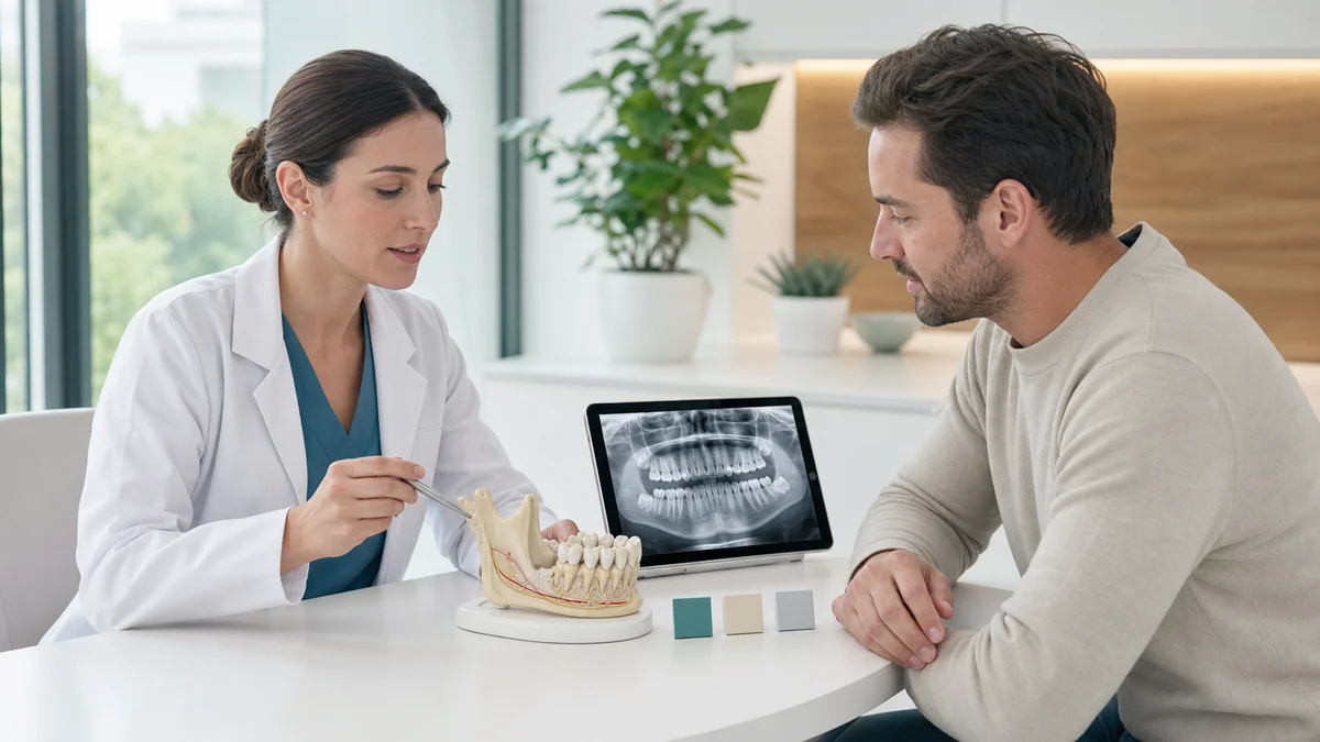
People searching for wisdom tooth removal alternatives may be trying to avoid surgery, nerve injury, recovery time or cost. A safe answer begins with the diagnosis. A healthy, disease-free wisdom tooth may be monitored. A high-risk lower tooth may be suitable for coronectomy. Local treatment can settle selected gum inflammation, but it may not prevent recurrence. A diseased tooth often still needs definitive care.
This guide explains what each option can and cannot do. It does not decide whether an individual tooth should remain, because that requires symptoms, examination, dental and medical history, and suitable imaging. If there is spreading swelling, difficulty breathing or swallowing, uncontrolled bleeding, fever with worsening dental symptoms, or severe illness, seek urgent professional help rather than comparing elective options online.
First Ask Whether the Wisdom Tooth Is Actually the Problem
Pain at the back of the jaw is not a diagnosis. A partly erupted wisdom tooth can develop inflamed gum around its crown, called pericoronitis, but similar discomfort may come from decay in the neighboring second molar, a cracked tooth, gum disease, a jaw-joint disorder, muscle pain, an ulcer or another condition. Removing a tooth that is not responsible may not resolve symptoms.
A dentist should ask when symptoms began, whether they recur, and whether there is swelling, bad taste, discharge, altered sensation, restricted mouth opening or difficulty swallowing. The examination evaluates the wisdom tooth, the tooth in front, surrounding gum, bite and soft tissues. A panoramic radiograph may show position, root form, decay, bone changes and proximity to anatomical structures. More detailed imaging is used only when it can change management.
That diagnostic step creates an important alternative: treat the condition that is actually present. If the second molar is the source and can be restored, its treatment may relieve pain without wisdom tooth surgery. If the symptoms are muscular or joint-related, an oral-surgery procedure may be inappropriate. This is not a loophole to preserve every wisdom tooth; it is accurate problem identification.
Wisdom Tooth Removal Alternatives at a Glance
| Finding or situation | Option to discuss | Main limitation |
|---|---|---|
| Disease-free, asymptomatic impacted tooth | Active monitoring | Future disease is still possible, so review and appropriate imaging cannot be abandoned. |
| First mild episode of gum inflammation around a partly erupted tooth | Professional local measures plus short-term self-care instructions | Symptoms may recur if the tooth remains difficult to clean or cannot erupt into a maintainable position. |
| Trauma from the opposing upper tooth onto inflamed gum | Assessment of the opposing tooth or bite | Adjusting or removing another tooth is appropriate only when trauma is confirmed and long-term function is considered. |
| Lower wisdom tooth requiring treatment with roots very close to the inferior alveolar nerve | Coronectomy in a suitable case | It is still surgery; retained roots can move, become infected or occasionally need later removal. |
| Pain caused by a restorable neighboring tooth | Treat the neighboring tooth | The wisdom tooth still needs its own assessment and may remain a risk to that restoration. |
| Medical condition or medicine makes elective timing less favorable | Coordinate, stabilize and schedule safely | Delay is not the same as avoiding indicated care, especially when infection is active. |
| Repeated infection, non-restorable decay, abscess, cyst or damage | Definitive treatment, often complete removal | Symptom control alone may allow disease to persist or worsen. |
The table is a conversation guide, not a diagnosis. Some options are alternatives to immediate extraction, while others change the surgical technique or treat a different source. Their value depends on why treatment was proposed in the first place.
1. Active Monitoring for a Disease-Free Wisdom Tooth
Monitoring is the clearest non-surgical option for a wisdom tooth that has no associated disease. The NICE guidance on wisdom tooth extraction recommends against prophylactic removal of pathology-free impacted third molars in the NHS. The NHS wisdom tooth removal page similarly explains that a wisdom tooth not causing problems will usually be left and checked at routine appointments.
Active monitoring means more than waiting for pain. Some disease can develop before it becomes obvious. A monitoring plan may include:
- Review of symptoms and any episodes of swelling or infection;
- Clinical examination of eruption, cleanliness and gum condition;
- Assessment of decay or periodontal damage on the second molar;
- Imaging when the clinician considers it justified;
- Clear instructions about symptoms that require an earlier visit;
- Reconsideration if health, access to care or treatment plans change.
Monitoring is most persuasive when the tooth is healthy and surgical benefit is uncertain. It is less likely to be appropriate when there is non-restorable decay, abscess, cystic change, resorption, damage to the adjacent tooth or repeated significant infection. A patient should ask the dentist to show what makes the tooth disease-free and what specifically will be watched.
2. Professional Local Care for Selected Pericoronitis
Pericoronitis is inflammation or infection of gum around a partially erupted tooth. Food and plaque can collect under the gum flap, and an opposing tooth may traumatize the tissue. A first episode does not automatically require removal. NICE states that a first episode, unless particularly severe, should not by itself be considered an indication for surgery, while repeated episodes can support removal.
The Scottish Dental Clinical Effectiveness Programme guidance prioritizes professional local measures such as debridement and irrigation where possible. The dentist may remove trapped material, assess whether the gum is being bitten, and give individualized cleaning and short-term symptom-care instructions.
Local care can be definitive if the tooth is still erupting into a cleanable, functional position and inflammation does not recur. It can also be temporary if there is insufficient space, persistent food trapping or repeated infection. The follow-up decision should consider eruption path and recurrence, not only whether symptoms improved for a few days.
3. Improve Access and Oral Hygiene Without Mistaking It for a Cure
Careful cleaning around a partly erupted tooth can reduce plaque and trapped food. A dental professional may demonstrate a small-headed or single-tufted brush and advise a rinse for a limited period when appropriate. The exact product must fit the patient’s health, allergies and other medicines.
Better hygiene is valuable, but it has limits. If the tooth remains covered by a deep flap, angled against the second molar or inaccessible, home care may not reach the disease-prone area. Aggressive probing, high-pressure irrigation or unapproved chemicals can injure tissue and should not be improvised.
Use hygiene as part of a reviewed plan rather than proof that extraction will never be needed. A good question is: can this tooth realistically become and remain cleanable, or are local measures only settling the current episode?
4. Manage Confirmed Trauma From the Opposing Tooth
Sometimes an upper tooth repeatedly bites the swollen gum over a lower wisdom tooth, worsening inflammation. SDCEP guidance notes that the opposing tooth may be assessed for adjustment or extraction where trauma is present and the tooth is unlikely to achieve useful future function.
This is not a generic shortcut. Removing or altering a sound opposing tooth changes the bite and must have a defensible clinical reason. The dentist should confirm contact, evaluate both third molars and consider whether the lower tooth can erupt. Simply choosing the easier tooth to remove without a complete assessment may trade one problem for another.
If trauma is one component of infection around a poorly positioned lower tooth, treating the upper tooth may reduce injury but may not eliminate food trapping or disease below the gum. Follow-up remains necessary.
5. Coronectomy for Selected High Nerve-Risk Lower Teeth
Coronectomy is the most important surgical alternative to complete lower wisdom tooth removal. The crown is removed while roots close to the inferior alveolar nerve are intentionally left in place. Its purpose is to reduce the risk of injuring the nerve that supplies sensation to the lower lip, chin, gums and teeth.
The updated University College London Hospitals coronectomy guidance describes monitoring, complete removal and coronectomy as options that may be discussed after specialist assessment. A two-dimensional radiograph is usually reviewed first; a cone-beam CT scan may be considered when more detailed information about the nerve relationship would change the decision.
Coronectomy is not suitable for every case. A tooth with decay or infection involving the roots may be unsuitable. During surgery, roots that become mobile may need to be removed. Retained roots can later migrate, erupt or become infected, and a minority of patients may need a second procedure. Coronectomy lowers a particular risk; it does not remove all surgical risk or eliminate follow-up.
Questions to ask a specialist include:
- What imaging signs suggest a close nerve relationship?
- Would additional imaging change management?
- Why is coronectomy suitable or unsuitable for this tooth?
- What happens if the roots move during the procedure?
- How will retained roots be reviewed afterward?
- Which symptoms could indicate infection or root migration?
- How would later root removal be managed if needed?
6. Treat a Different Confirmed Source of Pain
The second molar in front of a wisdom tooth can develop decay or gum damage, especially when the contact area is hard to clean. If that tooth is causing pain and can be restored, a filling, root canal treatment, periodontal care or another appropriate intervention may be discussed. The exact treatment follows the second molar’s diagnosis.
Treating the second molar is a genuine alternative only when it is the source and the wisdom tooth does not continue to threaten it. If the third molar’s angle creates an inaccessible defect, restoring the second molar without addressing the cause may produce a poor prognosis. A clinician should explain how both teeth affect the plan.
Jaw-joint and muscle conditions can also mimic third-molar pain. Pain with jaw movement, clenching or muscle tenderness requires appropriate assessment. An X-ray showing an impacted tooth does not prove that it causes every symptom.
7. Coordinate Timing When Medical Risk Can Be Improved
Elective oral surgery may need coordination when a patient has an unstable health condition, takes anticoagulant or antiplatelet medicine, is receiving treatment affecting bone or immunity, has had previous anesthesia problems, or is planning another major medical procedure. The safe response is coordinated care, not stopping prescribed medicine independently.
A dentist or oral surgeon may consult the prescribing clinician, choose an appropriate setting, modify the surgical plan or schedule treatment when risk is better controlled. An acute spreading infection can make delay unsafe, so timing decisions must distinguish elective risk reduction from urgent disease management.
Tell the team about all medicines and supplements, allergies, pregnancy, smoking or vaping, immune conditions, bleeding disorders and previous healing problems. A complete history can change both the choice and the setting of treatment.
8. Specialist Review and Modified Surgery
A referral to an oral surgeon does not create a non-surgical option, but it can create safer choices. The specialist can confirm whether treatment is needed, assess anatomy, discuss coronectomy and plan access, anesthesia and follow-up. A complex tooth that appears intimidating in general practice may have a clearer risk-management pathway after specialist review.
Modified surgical technique, sectioning the tooth, choosing local anesthesia with or without sedation, and selecting an appropriate care setting are not alternatives to removal itself. They are ways to deliver an indicated procedure. The patient should understand this distinction when comparing options.
A second opinion can be reasonable when the indication is unclear, the tooth is close to a nerve, or the choices have substantially different risks. Bring the existing images and treatment recommendation so the second clinician can evaluate the same question rather than repeat tests unnecessarily.
9. Short-Term Symptom Control While Awaiting Definitive Care
Pain control, a soft diet and clinician-directed local care may help while a patient awaits assessment or scheduled treatment. These measures can improve comfort, but they do not convert a diseased tooth into a healthy one. Repeatedly cycling through temporary remedies can allow decay, periodontal damage or infection to progress.
Medicine choice should reflect health history, pregnancy, allergies, kidney or liver disease, ulcers, asthma and interactions. Follow the label and professional advice; do not combine products with duplicate ingredients or exceed recommended doses. A pharmacist or clinician can help when suitability is uncertain.
Avoid placing aspirin, essential oils, concentrated antiseptics or caustic home remedies directly on the gum. These can burn tissue and obscure the clinical picture. Do not attempt to cut a gum flap, drain swelling or manipulate an impacted tooth at home.
Why Antibiotics Are Not a Routine Alternative to Removal
Antibiotics may be appropriate when there are signs of spreading or systemic infection, or for selected higher-risk patients after face-to-face assessment. They are not routinely needed for every painful wisdom tooth. The SDCEP guidance advises against antibiotics for pericoronitis unless there is spreading infection, systemic involvement, restricted opening associated with spread, or another relevant clinical reason.
NHS England’s urgent dental care guidance emphasizes that antimicrobials are an adjunct, not a substitute for appropriate local intervention. Antibiotics cannot correct an impacted position, remove non-restorable decay, clean under a persistent gum flap or eliminate a cyst.
Unnecessary antibiotic use can cause side effects and contribute to antimicrobial resistance. If a clinician prescribes one, take it as directed and still attend the planned dental review. Improvement does not prove that definitive treatment is unnecessary.
When Complete Removal May Still Be the Safest Choice
NICE lists pathological indications such as non-restorable decay, non-treatable pulpal or periapical disease, cellulitis, abscess, osteomyelitis, resorption, fracture, cyst or tumor, damage to adjacent structures and interference with other necessary surgery. Repeated or severe pericoronitis may also support removal after assessment.
In these situations, asking for alternatives should lead to an informed comparison, not a promise that surgery can always be avoided. The relevant comparison may be complete removal versus coronectomy, or prompt removal versus a short medically coordinated delay. Monitoring an active abscess as though it were a healthy tooth is not equivalent care.
Benefits of removal can include eliminating the diseased tooth and improving access to the second molar. Risks include pain, swelling, bleeding, infection, dry socket, injury to neighboring structures and altered sensation from nerve injury. Upper teeth may have sinus-related considerations. The balance is individual.
How to Build a Decision With Your Dentist
Ask the clinician to separate the indication from the technique. The indication explains why anything should be done. The technique explains how the selected treatment will be delivered. If these are blended together, a patient may hear “surgery” without understanding the disease, or hear “monitoring” without understanding the review plan.
A complete discussion should cover:
- The diagnosis and evidence on examination or imaging;
- The expected course if no procedure is performed now;
- Whether the tooth is disease-free, symptomatic or causing structural damage;
- Non-surgical measures and whether they are temporary or potentially definitive;
- Suitability for coronectomy if nerve proximity is important;
- Benefits and risks of complete removal;
- The follow-up required for each option;
- Urgent warning signs and whom to contact;
- Expected fees without treating an estimate as a fixed guarantee.
At Redent Klinik, an appropriate examination can organize these factors into a patient-specific plan. Patients seeking an assessment can use the Redent Klinik contact page and bring available radiographs, a medicine list and notes about previous episodes. A remote conversation can clarify questions, but it cannot replace clinical assessment or imaging when indicated.
After Choosing Monitoring: Make It an Active Plan
If the decision is to retain the tooth, agree on how it will be reviewed. The interval depends on eruption, cleanliness, age, neighboring teeth, prior symptoms and the clinician’s judgment. Routine imaging at arbitrary intervals is not automatically necessary; exposure should be justified by the information it may provide.
Maintain oral hygiene around the tooth as instructed and report new pain, swelling, food trapping, unpleasant taste, discharge or difficulty opening the mouth. A change in symptoms may call for earlier assessment. Monitoring should be revisited if the patient will lose access to care for a prolonged period or starts medical treatment that changes infection or surgical risk.
Keep a record of prior episodes and treatment. Recurrent pericoronitis can be underestimated when each episode is seen by a different urgent-care provider. Dates, severity, swelling and medicines help the regular dentist understand the pattern.
After Coronectomy or Removal: Recovery Is Still Surgery
Both coronectomy and complete removal create a surgical wound. Follow the treating team’s instructions on bleeding control, eating, cleaning, activity, smoking and medicines. Temporary pain, swelling, bruising and jaw stiffness can occur. Do not disturb the clot or surgical site with forceful rinsing or probing.
Contact the dental team for bleeding that does not stop as instructed, severe or worsening pain and swelling, fever, feeling unwell, pus, a worsening bad taste, or new altered sensation. Dry socket often presents as increasing pain several days after extraction and needs professional assessment. Severe swelling affecting breathing or swallowing requires emergency medical help.
After coronectomy, attend recommended reviews because retained roots may migrate. Movement is not automatically a complication, but exposure, infection or symptoms may require reassessment. The value of coronectomy depends partly on appropriate follow-up.
Frequently Asked Questions About Wisdom Tooth Removal Alternatives
What are the safest wisdom tooth removal alternatives?
The safest option depends on the diagnosis. Active monitoring can be appropriate for a disease-free tooth. Local professional treatment may manage a selected first episode of pericoronitis. Coronectomy may reduce nerve-injury risk for a suitable lower tooth close to the inferior alveolar nerve. No option is universally safest.
Can I keep an impacted wisdom tooth forever?
Possibly, if it remains disease-free and a dentist considers monitoring appropriate. Retention still needs review because decay, gum damage, cystic change or damage to the second molar can develop. Lack of pain alone does not prove that the tooth is healthy.
Can mouthwash replace wisdom tooth removal?
No mouthwash changes the angle of an impacted tooth or cures non-restorable decay, an abscess or a cyst. A clinician-directed rinse may support short-term hygiene during gum inflammation, but it is not a universal definitive treatment. Persistent or recurrent symptoms need dental reassessment.
Is coronectomy better than complete wisdom tooth removal?
Coronectomy can be preferable in a selected lower tooth when complete removal carries an elevated inferior alveolar nerve risk. It is unsuitable for some teeth and can require later review or root removal. Complete removal may be more appropriate when root disease or other contraindications are present.
Can antibiotics make wisdom tooth surgery unnecessary?
Antibiotics may help selected spreading or systemic infections, but they do not correct impaction or remove the underlying source. They are generally an adjunct to professional care, not a replacement. Repeated prescriptions without a definitive plan can delay necessary treatment and contribute to resistance.
Can the gum over a wisdom tooth be removed instead?
Soft-tissue treatment may be considered in selected cases, but suitability depends on whether the tooth can erupt into a functional, cleanable position and whether inflammation is likely to recur. Removing gum alone does not solve decay, poor angulation, cystic disease or damage to a neighboring tooth.
Can braces create space instead of wisdom tooth removal?
Orthodontic treatment is not a routine substitute for managing a diseased wisdom tooth. In unusual comprehensive plans, tooth movement or eruption management may affect third-molar decisions. That requires an orthodontist and dentist or surgeon to evaluate the whole bite, not an assumption that braces can rescue every impacted tooth.
Should I get a second opinion before wisdom tooth surgery?
A second opinion can be useful when the indication is unclear, the tooth is close to a nerve or options have meaningfully different risks. Seek urgent care rather than delaying if there is spreading infection, airway concern or uncontrolled symptoms. Bring existing images to reduce unnecessary duplication.
Does sedation count as an alternative to removal?
No. Sedation changes how anxiety and awareness are managed during treatment; it does not change the diagnosis or remove the need for a procedure. Local anesthesia, sedation and general anesthesia have different indications and risks that the treating team should explain.
What happens if I choose no treatment?
For a disease-free tooth, no immediate procedure plus active monitoring may be appropriate. For a diseased tooth, choosing no treatment can allow pain, infection or structural damage to continue. Ask the dentist to describe the likely course, warning signs and review plan for your specific findings.
Final Answer: The Best Alternative Must Match the Diagnosis
The most credible wisdom tooth removal alternatives are active monitoring for disease-free teeth, professional local care for selected pericoronitis, treatment of another confirmed pain source, and coronectomy for some lower teeth with high nerve proximity. Specialist review and medical coordination can also reduce risk or improve timing.
These options are not interchangeable. Temporary pain control and antibiotics do not reliably replace definitive treatment when there is non-restorable decay, abscess, cyst, repeated infection or damage. Ask the clinician to show the diagnosis, explain the likely course with and without intervention, and define the follow-up for every proposed alternative.
Official Sources and Further Reading
- NICE: Guidance on the extraction of wisdom teeth
- NHS: Wisdom tooth removal
- University College London Hospitals: Wisdom tooth surgery and coronectomy
- Scottish Dental Clinical Effectiveness Programme: Acute pericoronitis
- NHS England: Urgent and non-urgent dental care
- British Association of Oral Surgeons: Coronectomy patient information
- American Dental Association
- World Health Organization: Oral health fact sheet
Sources reviewed July 2026. This article is general education and does not replace examination, diagnosis, imaging or an individualized consent discussion.