
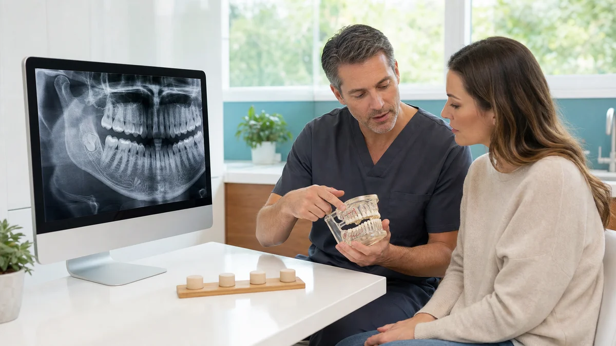
Quick answer: A useful wisdom tooth removal comparison starts with diagnosis, not technique. A healthy, trouble-free tooth may be monitored; disease can support removal. Erupted teeth may need a simple extraction, while impacted teeth can require surgery. For a lower tooth close to a nerve, coronectomy may sometimes reduce nerve risk. Examination and suitable imaging determine the safest route.
A wisdom tooth removal comparison is not simply a contest between a “quick” extraction and an operation. The first decision is whether intervention is justified at all. The next decisions concern access to the tooth, the relationship of its roots to nerves or the sinus, the appropriate form of anaesthesia, the setting, aftercare and the plan if healing does not follow the expected course. Two teeth that look similar in a mirror can require very different management.
This guide provides a structured way to discuss those choices with a dentist or oral surgeon. It does not diagnose an individual tooth and does not promise that one approach will be painless, complication-free or suitable for everyone. A clinical examination, medical and dental history, and imaging selected for a clear clinical purpose remain essential. The aim is informed consent: understanding why a recommendation has been made, what reasonable alternatives exist, and how follow-up will work.
1. Begin the Wisdom Tooth Removal Comparison With “Monitor or Treat?”
Wisdom teeth, also called third molars, may erupt fully, emerge only partly, remain covered by gum, or stay within bone. Position alone is not the same as disease. A tooth can be impacted without causing current symptoms, while another can be partly erupted and repeatedly inflame the surrounding gum. The decision therefore needs evidence from symptoms, examination and suitable radiographs rather than a rule that every wisdom tooth should be removed.
The National Institute for Health and Care Excellence advises against prophylactic removal of pathology-free impacted third molars in its NHS guidance. It identifies evidence of pathology, such as unrestorable decay, non-treatable pulpal or periapical disease, abscess, cystic change, resorption or other defined problems, as reasons that can support surgery. The American Dental Association’s patient guidance likewise describes pain, infection, cysts, damage to neighbouring teeth, gum disease and non-restorable decay as findings that may lead to removal.
When monitoring may be reasonable
Monitoring may be discussed when a tooth is currently free from relevant disease, does not damage the second molar, can be assessed reliably and the patient can attend reviews. Monitoring is an active plan, not forgetting that the tooth exists. The clinician should explain what will be checked, how often reassessment is appropriate for that patient, and which symptoms should trigger an earlier appointment.
- There is no current pain, swelling, infection, cystic change or other treatment indication.
- The tooth and the neighbouring molar can be examined and kept acceptably clean.
- Available imaging does not show a problem requiring intervention.
- The patient understands that future disease remains possible and agrees to review.
When removal deserves a focused discussion
Removal can become more reasonable when there is a diagnosed problem or a pattern of recurrence. Examples include decay that cannot predictably be restored, repeated inflammation around a partly erupted tooth, an abscess, periodontal damage on the neighbouring molar, resorption, a cyst or interference with another necessary operation. A first mild episode of pericoronitis is not automatically the same as repeated or severe disease; the clinical history matters.
Symptoms alone also need interpretation. Jaw pain can come from several sources, and removing a wisdom tooth that is not responsible may not resolve it. Conversely, absence of pain does not prove the absence of disease. This is why the diagnosis and its evidence should appear clearly in the treatment record and consent conversation.
2. Decision Table: Four Main Management Routes
The following table is a conversation aid, not a self-selection tool. “Best” means the option whose expected benefit is proportionate to its risks for the individual tooth and patient.
| Route | When it may enter the discussion | What it involves | Important limitation or risk | Follow-up question |
|---|---|---|---|---|
| Active monitoring | No current relevant pathology and reliable review is possible | Clinical checks, hygiene support and imaging only when clinically justified | Future problems can still develop | What change should bring me back sooner? |
| Simple extraction | Tooth is sufficiently erupted and accessible | Removal through the socket, usually without a gum flap or bone removal | “Simple” describes access, not a guarantee of an easy recovery | What could make the plan change during treatment? |
| Surgical extraction | Tooth is partly or fully impacted, broken down, or not safely accessible by a simple route | May include a gum incision, controlled bone removal, tooth sectioning and stitches | Nerve, sinus, adjacent-tooth, dry-socket, infection and bleeding risks vary by case | Which anatomical feature creates my main risk? |
| Coronectomy | Selected lower wisdom tooth whose roots are very close to the inferior alveolar nerve | Crown is removed while suitable roots are intentionally retained | Not suitable for every tooth; retained roots may migrate or rarely need later care | Why is this safer than complete removal in my case? |
The route can change if new information appears. For example, an apparently accessible tooth may fracture, or imaging may show a nerve relationship that warrants specialist review. A sound consent process covers foreseeable changes and explains who will make decisions if the original plan cannot be completed as expected.
3. Simple Extraction Versus Surgical Extraction
What makes an extraction “simple”?
A simple extraction is generally possible when enough of the crown is visible and instruments can safely mobilise the tooth through its socket. Local anaesthetic is commonly used. The absence of an incision or bone removal can mean less tissue manipulation, but the experience still depends on root shape, bone density, inflammation, access and the patient’s medical factors. The word “simple” should never be interpreted as zero risk.
The clinician should still assess the neighbouring tooth, root anatomy and whether the tooth could communicate with the maxillary sinus or lie near a nerve. Even an erupted wisdom tooth may be heavily decayed, fragile or difficult to access. A backup surgical plan can therefore be appropriate.
What changes in surgical removal?
Surgical extraction creates controlled access. The surgeon may lift a gum flap, remove a limited amount of surrounding bone, divide the tooth into sections and close the area with dissolving or removable stitches. Sectioning is not evidence that something has gone wrong; it can reduce the amount of force or bone removal needed. The exact sequence should be tailored to anatomy rather than repeated as a standard package.
The NHS wisdom tooth removal overview describes these possible steps and explains that local anaesthetic is usual, with sedation or general anaesthesia considered in selected circumstances. Complexity, anxiety, medical history, service capability and the number of teeth being treated all influence the plan.
4. Full Removal Versus Coronectomy Near the Lower-Jaw Nerve
Lower wisdom tooth roots can lie close to the inferior alveolar nerve, which supplies sensation to the lower lip and chin. The lingual nerve, which contributes sensation to the tongue, is another relevant structure. Temporary altered sensation is a recognised complication of lower wisdom tooth surgery; permanent change is less common but important enough to discuss clearly. Individual risk cannot be estimated responsibly from a generic internet percentage.
How coronectomy changes the trade-off
Coronectomy removes the crown and leaves suitable roots in place, reducing deliberate manipulation around a high-risk nerve relationship. University College London Hospitals describes it as an option that may be offered when complete removal would carry an increased nerve-injury risk. It is not a shortcut for every impacted tooth.
The roots should be suitable for retention, and the surgeon must consider infection, tooth vitality, mobility and other clinical factors. Retained roots can move over time; this may be harmless, may make later removal safer, or may occasionally create a need for another procedure. Patients need to understand the follow-up plan and the possibility that conversion to full removal could become necessary during surgery if the roots become mobile or circumstances change.
Questions that make the comparison useful
- Which radiographic signs suggest that the roots and nerve are close?
- Would additional imaging change the treatment route or only add detail?
- Is the tooth suitable for coronectomy, and what findings would rule it out?
- How will retained roots be reviewed, and who provides care if symptoms appear later?
5. Panoramic X-Ray Versus CBCT
Imaging should answer a clinical question. A panoramic radiograph often shows the tooth’s position, root development, adjacent teeth and broad relationship to the mandibular canal or maxillary sinus. It can support diagnosis and planning without automatically exposing a patient to three-dimensional imaging.
Cone-beam computed tomography, or CBCT, can show anatomy in three dimensions. However, more detail is not automatically better care. The US Food and Drug Administration states that dental CBCT generally delivers more radiation than conventional dental X-rays and should be used only when necessary to provide clinical information unavailable from other modalities. The useful question is therefore: will CBCT change the management decision, technique or consent discussion?
A proportionate imaging sequence
A clinician can start with history, examination and existing images. If a new panoramic image is justified, it may be enough. When two-dimensional signs suggest a close nerve relationship and precise localisation could alter full removal, coronectomy or referral, CBCT may be considered. Exposure settings and field of view should be appropriate to the clinical need. Patients should also bring recent images to avoid needless duplication where the files remain diagnostically adequate.
6. Local Anaesthetic, Sedation or General Anaesthesia
Anaesthesia choice should match the operation, patient and clinical setting. More intensive anaesthesia is not automatically more comfortable overall or more appropriate. Each option changes preparation, monitoring, escort requirements, recovery and risk.
Local anaesthetic
Local anaesthetic numbs the treatment area while the patient remains awake. Pressure and movement may still be perceived, but sharp pain should not be accepted as inevitable. The patient and clinician should agree on a stop signal, and additional anaesthetic can be considered if needed. Local anaesthetic often allows a simpler recovery from the anaesthetic itself, although surgical recovery is still determined by the operation.
Conscious sedation
Sedation may be discussed for significant anxiety, a longer procedure or other selected needs. The patient is usually responsive but more relaxed; memory of the procedure may be reduced. The method, fasting rules, monitoring, escort and post-treatment restrictions depend on local protocols and the drugs used. A responsible provider gives written instructions before the appointment, not after the sedative has been administered.
General anaesthesia
General anaesthesia makes the patient unconscious and requires a suitably equipped setting and trained team. It may be appropriate for particular clinical situations, but it brings its own assessment and recovery considerations. It should not be presented as a premium upgrade. The indication, alternatives and anaesthetic risks should be discussed separately from the surgical risks.
Medical history can change all three routes. Allergies, pregnancy, sleep apnoea, heart or lung conditions, previous anaesthetic problems, anticoagulants, antiplatelet medicines and other prescriptions or supplements should be disclosed. Patients should not stop prescribed medication unless the responsible clinician has provided an individual plan.
7. Lower Wisdom Teeth Versus Upper Wisdom Teeth
The jaw changes the risk map. For lower wisdom teeth, the relationship to the inferior alveolar and lingual nerves often dominates planning. For upper wisdom teeth, the maxillary sinus and the bone behind the tooth are relevant. This does not mean all lower teeth are harder or all upper teeth are easy. Root form, eruption, mouth opening, adjacent restorations and local anatomy can reverse that expectation.
Lower-jaw considerations
In addition to nerve risk, a lower tooth may be angled against the second molar, partly covered by gum or surrounded by dense bone. The clinician should inspect the second molar for decay, periodontal pocketing or resorption because treating only the wisdom tooth does not automatically restore neighbouring structures. Restricted mouth opening and postoperative swelling can be more noticeable after deeper surgery.
Upper-jaw considerations
Upper roots may lie near the maxillary sinus. A communication between mouth and sinus can occur after removal and may require specific instructions or further treatment. Rarely, a tooth or root can displace, or the bone behind the molar can fracture. These possibilities should be placed in context by the treating clinician rather than used to frighten a patient.
8. One Tooth Versus Several in the Same Session
Removing several indicated teeth in one session can consolidate anaesthesia, time away from work and the main recovery period. It can also mean more treatment at once, broader swelling and greater reliance on support during the first days. Treating one side first may preserve a more comfortable chewing side and reveal how the individual heals, but it requires another appointment and recovery period if the remaining teeth still need care.
The decision should not be driven by package pricing. Each tooth needs its own diagnosis and risk assessment. A symptom-free tooth should not be added automatically merely because another tooth requires surgery. Conversely, when several teeth have clear indications and the anaesthetic plan is appropriate, staged care is not inherently safer. The consent record should identify exactly which teeth are planned and why.
9. General Dentist Versus Oral Surgeon and Treatment Setting
Many erupted or straightforward teeth can be managed by an appropriately trained dentist. Deep impaction, a high nerve or sinus risk, limited access, significant medical complexity, a need for advanced sedation or uncertainty in diagnosis can support referral to an oral surgeon or hospital service. Referral is a risk-management decision, not a sign that a dentist has failed.
Ask who will actually perform the procedure, their relevant training, where sedation or general anaesthesia will occur, and how emergencies are managed. The setting should have suitable monitoring, infection-control processes, imaging access and a clear transfer pathway. These fundamentals matter more than luxurious surroundings.
Records to collect before accepting a plan
- The diagnosis and indication for each tooth being treated.
- Copies of relevant X-rays or CBCT data and the imaging report where applicable.
- The planned technique, anaesthesia and any realistic alternative.
- Personalised material risks, including the main nerve or sinus concern.
- Written preparation, medication and aftercare instructions.
- A named contact route for urgent and routine postoperative questions.
10. Recovery Comparison: What Actually Changes
Recovery is influenced by depth, duration, tissue manipulation, infection, number of teeth, smoking or vaping, general health and adherence to instructions. A simple extraction often involves less surgical trauma than a deep bony impaction, but no clinician can guarantee an exact pain level or return-to-work date. Plan some flexibility rather than relying on a universal timeline.
The first phase of healing
A stable blood clot protects the socket. Some oozing, soreness, swelling, bruising and jaw stiffness can occur. Written instructions commonly cover pressure with gauze, food texture, oral hygiene, activity and pain relief. Vigorous rinsing, disturbing the socket and smoking can interfere with healing. Follow the treating team’s instructions because they may differ according to the operation and medical history.
Dry socket, or alveolar osteitis, occurs when the protective clot is lost or breaks down. Pain may intensify after initially seeming manageable and can radiate toward the ear or temple. It is not simply “normal pain that must be tolerated”; the dental team can assess and provide local care. Antibiotics do not replace assessment and are not a routine solution for every painful socket.
Medication and antibiotic decisions
Pain relief should be personalised around allergies, stomach, kidney, liver or cardiovascular conditions, pregnancy and other medicines. Patients should avoid combining products with overlapping ingredients. Antibiotics are not automatically required for every wisdom tooth extraction. Their use depends on diagnosis, systemic signs, individual risk and local guidance. Taking leftover antibiotics or someone else’s prescription can delay appropriate care and contributes to antimicrobial resistance.
When the recovery needs urgent review
Contact the treating service urgently if bleeding does not stop with the measures provided, pain or swelling is severe or worsening, or there is a bad taste together with fever or feeling unwell. Seek emergency help for breathing difficulty, rapidly spreading swelling, collapse or another severe systemic reaction. New or persistent numbness should also be reported so it can be documented and reviewed, even when early observation is the initial plan.
11. Travelling for Wisdom Tooth Surgery
Travel adds a second comparison: the procedure itself and the continuity of care around it. A compressed itinerary may leave little time for swelling to develop, for a socket to be reviewed or for a complication to be treated by the original team. The lowest quoted treatment fee does not represent the full decision if imaging, sedation, medicines, accommodation, changed flights or local follow-up are excluded.
Before travelling, obtain a written plan showing which teeth are being considered and whether a final decision depends on examination or new imaging. Ask how long the clinic recommends staying nearby, who can review bleeding or severe pain outside normal hours, and how records will be shared with a home dentist. Airlines and insurers may have their own requirements; these should be checked directly rather than inferred from a dental estimate.
Patients considering care in Istanbul can review Redent Klinik’s English-language clinic overview and use the contact page to request an individual assessment. Photos or an existing panoramic image can support an initial conversation, but they cannot replace an in-person examination or guarantee the final technique.
12. A Nine-Question Consultation Checklist
A high-quality wisdom tooth removal comparison should leave the patient able to explain the recommendation in their own words. Bring this checklist to the consultation and note the answers for each tooth:
- What is the diagnosis, and what evidence shows that treatment is needed now?
- What are the likely consequences of active monitoring instead?
- Is a simple extraction realistic, or is surgical access expected?
- For a lower tooth near the nerve, has coronectomy been considered and why?
- What imaging is necessary, and how will it change management?
- Which anaesthetic option is proportionate to my procedure and health?
- What are my most relevant risks rather than only generic complications?
- Who will provide routine and urgent follow-up, especially if I travel?
- What could cause the plan to change before or during treatment?
If the answers remain vague, it is reasonable to ask for clarification or a second opinion before non-urgent surgery. Informed consent is a process, not a signature obtained minutes before treatment.
13. Frequently Asked Questions About Wisdom Tooth Removal Comparison
Does every impacted wisdom tooth need to be removed?
No. Impaction describes position, not necessarily disease. A pathology-free tooth may be monitored when examination and review are reliable. Removal becomes more relevant when there is a defined problem, meaningful risk to a neighbouring structure or recurrent disease. The reason should be documented for each tooth rather than assumed from age or impaction alone.
Is simple extraction always safer than surgical extraction?
Not automatically. A simple route is appropriate only when the tooth can be removed with controlled access. Persisting with it when visibility or access is poor can create unnecessary force. A planned surgical approach may provide safer control for an impacted or fragile tooth. The correct comparison is between techniques suitable for that anatomy.
Is coronectomy better than complete wisdom tooth removal?
Coronectomy can be valuable for selected lower wisdom teeth whose roots are very close to the inferior alveolar nerve. It reduces root manipulation but intentionally leaves tissue that may migrate or occasionally require later care. It is not suitable when root retention would be unsafe. A surgeon must assess eligibility.
Do I need CBCT before wisdom tooth surgery?
Not routinely. A conventional panoramic image may provide enough information. CBCT is considered when three-dimensional detail is expected to change diagnosis, technique, referral or consent. Because it generally uses more radiation than conventional dental radiography, the examination should have a specific question and an appropriately limited protocol.
Is general anaesthesia the best choice for four wisdom teeth?
There is no universal best anaesthetic based only on the number of teeth. Complexity, anxiety, medical history, treatment duration, local capability and patient preference all matter. Local anaesthetic, sedation and general anaesthesia have different preparation, monitoring and recovery requirements. The provider should explain why the proposed level is proportionate.
Can a wisdom tooth be removed during active infection?
Sometimes definitive treatment proceeds, while in other situations drainage, supportive care or control of systemic infection comes first. Mouth opening, swelling, airway risk, medical status and access all affect timing. Antibiotics alone do not remove the source. Facial swelling, fever, difficulty swallowing or breathing requires prompt professional assessment.
Will removing wisdom teeth prevent crowding?
Crowding has multiple causes, and removal should not be promised as a guarantee that teeth will remain straight. If orthodontic treatment is planned, the orthodontist and surgeon should explain the specific purpose of removal in that plan. Retainers and long-term tooth movement need a separate discussion.
How long does recovery take?
There is no single reliable duration. Many people resume light activities relatively soon, while deeper surgery, multiple teeth or complications can extend recovery. Swelling and jaw stiffness can temporarily worsen before improving. The treating team should give a case-specific range and clear triggers for review rather than promise a fixed day of recovery.
Can I fly immediately after removal?
Do not assume that flying immediately is suitable, especially after upper wisdom tooth surgery near the sinus, sedation or a complicated extraction. The operating clinician must advise based on anatomy and recovery, while the airline and insurer set their own rules. A travel plan should include time for review and a route to urgent care.
What is the most important result of a wisdom tooth removal comparison?
The most important result is a defensible, individual plan: a clear diagnosis, a proportionate technique, understood alternatives, personalised risks and reliable follow-up. Choosing the most aggressive or most expensive option is not the goal. Choosing an approach whose expected benefit justifies its burden and risk is.
14. The Practical Bottom Line
A sound wisdom tooth decision starts by separating position from pathology. Monitoring can be responsible care when a tooth is healthy and review is reliable. When removal is justified, access determines whether a simple or surgical technique is realistic; nerve proximity may make coronectomy relevant; and the upper jaw introduces sinus-specific considerations. Imaging and anaesthesia should be selected because they change safety or care, not because they make a package appear more advanced.
The patient should leave with written preparation and aftercare, copies of relevant records, an urgent contact route and a clear understanding that the plan can evolve with new findings. This structure protects quality whether treatment occurs close to home or abroad.
Sources
- NICE: Guidance on the extraction of wisdom teeth, recommendations
- NHS: Wisdom tooth removal
- American Dental Association and ADA MouthHealthy: Wisdom teeth
- American Association of Oral and Maxillofacial Surgeons: Management of impacted third molar teeth
- University College London Hospitals: Advice for patients considering coronectomy
- US Food and Drug Administration: Dental cone-beam computed tomography
- World Health Organization: Oral health fact sheet